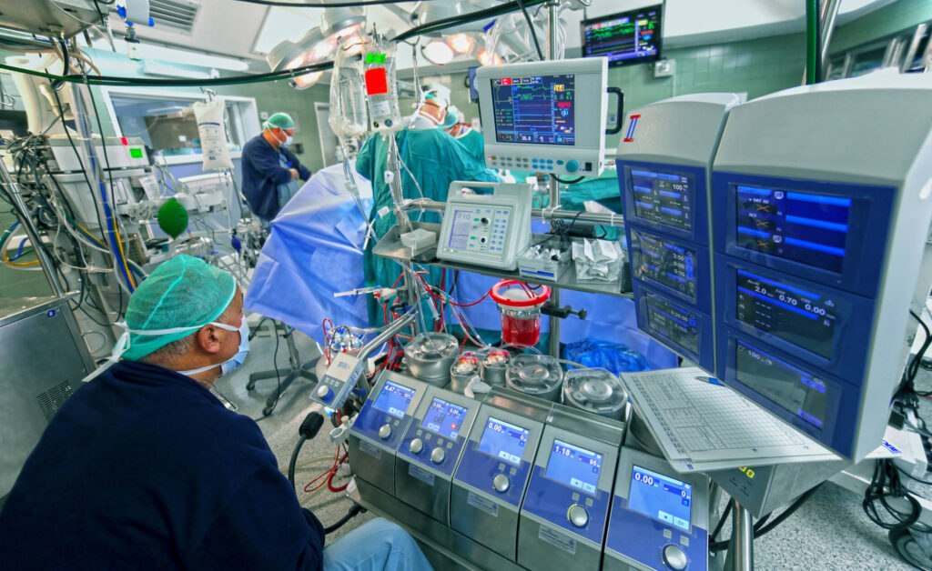
In recent years, the use of computers and machines in surgery has gained widespread attention and adoption. Some of the benefits include increased accuracy, decreased duration, and minimized invasiveness. As computer hardware continues to miniaturize, the deployment of robotic surgery inside the body has become more feasible. Application domains include eye and ENT surgery, cardiac surgery, laparoscopy, and neurosurgery. The development of these internal surgical robots can be modeled across five generations of research [1]. 

The “zeroth” generation is laparoscopy, involving camera-aided surgery through a small incision. Although traditional laparoscopy does not involve robots, it inspired the first generation of surgical robots: stereotaxic robots. These robots compute precise positions for each step in a surgical procedure, albeit without a visual aid like in laparoscopy. The second generation involved rigid dexterous robots, such as precise robotic arms, to carry out surgery. One notable example is the “da Vinci” platform, currently run by Intuitive Surgical Inc. Then came the third generation of flexible robots, where a robot could enter the human body and navigate through vessels and orifices. Up through this point, these technologies required a physical connection to some exterior device (power supply, computer, etc.). The fourth generation is the untethered microsurgeon, a free-floating device — for example, a capsule endoscope. 

Recent research on internal surgery focuses on third- and fourth-generation robots. Bruns et al. 2020 developed a tethered robot for inserting electrode arrays for cochlear implants, which are housed inside the delicate inner ear [2]. Empirical results show that manual insertions produce highly variable exerted forces, whereas their robot could alleviate much of these variations. In the automated process, the only human involvement is a button, which the surgeon holds to run a precomputed trajectory, allowing for a quick deactivation in the event of an emergency. 

Heunis et al. 2020 presented the ARMM system for a specific type of robotic surgery: positioning endovascular catheters [3]. The typical procedure involves inserting a catheter in the groin and using X-ray imaging to monitor the catheter’s progress through the body. The patient risks trauma during the positioning process, while both patient and clinician must be exposed to X-rays for some time. The ARMM system computes a trajectory from a CT scan, navigates a tethered magnet through the arterial tree, and monitors the catheter’s progress in real-time using ultrasound. Leclerc et al. 2020 designed an untethered “magnetic swimmer” that, when subjected to a magnetic field, rotates and swims within the bloodstream [4]. One application is the treatment of acute pulmonary embolism. In a setting where a timely response with minimal side effects is essential, treatments such as anticoagulants or thrombolysis are inefficient, invasive, or dangerous. Instead, the magnetic swimmer can navigate the body’s blood vessels (even the chambers of the heart), taking advantage of its rotating motion and helical design to abrade the embolism. 

The previous three projects use some form of custom electromagnets to control their respective robots. Erin et al. 2020 use magnetic fields generated by MRI devices to steer and control a robot with five degrees-of-freedom [5]. The cylindrical robot contains an air gap, inside which one can install cameras, lasers, or other components to achieve targeted yet minimally-invasive surgical procedures. While the magnetic swimmer is designed for blood vessels, the robot designed by Erin et al. is intended for organs. 

As the research progresses, the cost and size of fourth-generation surgical robots are likely to decrease, while their accuracy, capabilities, and usage increase. Some identified challenges include miniaturization to the micrometer or nanometer scale, propulsion beyond electromagnets, visual feedback for the supervising surgeon, and social acceptability [1]. 

References 

[1] C. Bergeles. From Passive Tool Holders to Microsurgeons: Safer, Smaller, Smarter Surgical Robots. IEEE Transactions on Biomedical Engineering 61.5, 1565-1576 (2014). 

[2] T. L. Bruns et al. Magnetically Steered Robotic Insertion of Cochlear-Implant Electrode Arrays: System Integration and First-In-Cadaver Results. IEEE Robotics and Automation Letters (2020). Presented at the 2020 IEEE International Conference on Robotics and Automation (ICRA). 

[3] C. M. Heunis et al. The ARMM System – Autonomous Steering of Magnetically-Actuated Catheters: Towards Endovascular Applications. IEEE Robotics and Automation Letters (2020). Presented at the 2020 IEEE International Conference on Robotics and Automation (ICRA). 

[4] J. Leclerc et al. Agile 3D-Navigation of a Helical Magnetic Swimmer. IEEE International Conference on Robotics and Automation (ICRA 2020). 

[5] O. Erin et al. Towards 5-DoF Control of an Untethered Magnetic Millirobot via MRI Gradient Coils. IEEE International Conference on Robotics and Automation (ICRA 2020). 

Categories
Uncategorized

The Anatomy of an N-95 Mask

An N95 respirator is a respiratory protective device designed to achieve a very close facial fit and very efficient filtration of airborne particles. The ‘N95’ designation means that when subjected to careful testing, the respirator blocks at least 95 percent of very small (0.3 micron) test particles. There are also N99 and N100 respirators (N100s stop at least 99.97% of particles from entering) (1). Legitimate surgical respirators must be approved by both the FDA and the NIOSH (the National Institute for Occupational Safety and Health). Some N95s feature exhalation valves, which help the wearer to breathe more easily. It is important to note that not all N95 respirators are designed for medical applications; some are manufactured for industrial use. Medical N95s are single-use products regulated as class II products under the FDA and NIOSH (1). Although the demand for N95s has greatly increased during the current coronavirus (COVID-19) pandemic, the CDC recommends that N95 masks must continue to be reserved for health care workers and other medical first responders (2). For members of the general public, the CDC recommends the use of simple cloth face coverings when in a public setting to slow the spread of the virus (2). 

An N95 respirator consists of multiple layers of nonwoven fabric, often made from polypropylene. The two outward protective layers of fabric, covering the inside and outside of the mask, are between 20 and 50 g/m2 in density and act as protection against the outside environment, as well as a barrier to anything in the wearer’s exhalations (1). Between the two aforementioned ‘outer’ layers is a pre-filtration layer, which can be as dense as 250 g/m2, and the filtration layer. The pre-filtration layer is usually needled, nonwoven fabric. Needled nonwoven material is created by sending barbed needles repeatedly through the fabric to hook fibers together, in order to increase the cohesiveness of the material. The pre-filtration layer is then run through a hot calendering process, in which plastic fibers are thermally bonded by running them through high pressure heated rolls. This makes the pre-filtration layer thicker and stiffer, so it can be molded to form the desired shape and stay in that shape as the mask is used (1). The last layer is a high efficiency, melt-blown, polarized nonwoven material, which determines the filtration efficiency. Meltblowing is a process in which multiple machine nozzles use air to spray threads of melted synthetic polymers onto a conveyor. As the conveyor continues, the threads build up and bond by themselves as they cool, thereby creating the fabric. Sometimes, melt-blown fabric is also thermally bonded to add strength and abrasion resistance, although the material then begins to lose some of its fabric characteristics (1). The full respirators are made through converting machinery, which combines the layers through ultrasonic welding and adds straps and metal strips to adjust the mask over the user’s nose. The respirators are then sterilized as a last step before being shipped (1). 

On April 3, drastic shortages of N95 masks led the FDA to allow imports of similar masks from China. These masks, known as KN95, were never tested by American regulators, but were required to be vetted by an accredited test laboratory, which could be outside the United States, showing that they met the standards of the CDC. As an additional safeguard, the CDC and the FDA initiated their own review to make sure the masks met these performance standards. In tests of about 11 masks that the FDA had authorized to be sold to American hospitals, seven flunked (3). One mask removed as little as 24 to 35 percent of particles, according to a CDC test on April 15. Others just barely missed the cut, such as masks from DaddyBaby, a company in Fuzhou, China, that typically makes diapers. Those masks removed between 91 percent and 93.5 percent of particles. The health agencies also tested some KN95 masks that did not have agency approval. One of these, the F.D.A. said, blocked roughly 1 percent of the particles. On May 7, the FDA barred more than 65 of 80 previously authorized manufacturers in China from exporting these KN95 masks to the United States for medical use (3).  

Nevertheless, there is no need to worry about the availability of N95 masks in the US, as both Honeywell and 3M have promised to ramp up their production of these vital respirators. As of March 23, 3M has doubled its global production of N95 masks to about 100 million a month, and is planning to invest in new equipment to push annual mask production to 2 billion within 12 months (4). On March 22, Chief Executive Officer Mike Roman said in a news release that 3M had sent 500,000 respirators to hard-hit Seattle and New York City, and that it was ramping up production of hand sanitizers and disinfectants as well. Two days later, Roman said 3M would work with Ford Motor Co. to produce powered air purifying respirators, waist-mounted devices that blow air into helmets that shield wearers. Honeywell is also increasing N95 production, saying it will hire at least 500 people to expand capacity at a facility in Rhode Island (4). 

References 

  1. Henneberry, B. (n.d.). How to Make N95 Masks. Retrieved from https://www.thomasnet.com/articles/plant-facility-equipment/how-to-make-n95-masks/ 
  2. Center for Devices and Radiological Health. (n.d.). N95 Respirators and Surgical Masks (Face Masks). Retrieved from https://www.fda.gov/medical-devices/personal-protective-equipment-infection-control/n95-respirators-and-surgical-masks-face-masks#s3 
  3. Nicas, J., & Kaplan, S. (2020, May 7). F.D.A. Bans Faulty Masks, 3 Weeks After Failed Tests. Retrieved from https://www.nytimes.com/2020/05/07/health/masks-banned-n95-coronavirus.html 
  4. Gruley, B., & Clough, R. (2020, March 25). How 3M Plans to Make More Than a Billion Masks By End of Year. Retrieved from https://www.bloomberg.com/news/features/2020-03-25/3m-doubled-production-of-n95-face-masks-to-fight-coronavirus 

The incidence and prevalence of liver disease (particularly alcoholic liver disease and hepatitis C) is increasing in the developed world. In the UK, in 2006, 4450 people died from alcoholic liver disease, and deaths are increasing by 7% per year (1). Liver disease can be acute or chronic. Common causes of chronic liver disease are viral hepatitis (hepatitis B and C), autoimmune disease, and alcoholic liver disease. In the USA and the UK, acetaminophen overdose is the most common cause of acute hepatic failure, while worldwide it is viral hepatitis (2). Despite the diversity of the causes of liver disease, the outcome after anesthesia and surgery depends more on the degree of liver impairment than the actual cause. Patients with end-stage liver disease are at significant risk of morbidity and mortality after anesthesia and surgery, and thus present unique challenges for the anesthesiologist.  

Preoperative assessment of patients with liver disease should focus on the extent of liver dysfunction and extra-hepatic complications (1). A full blood count will detect anemia, thrombocytopenia, or raised white cell count if infection is present. Prothrombin time (PT) is a useful indicator of hepatocellular function and is used as a prognostic indicator in acute liver failure and after surgery in patients with chronic liver disease. However, PT may be elevated independent of liver function in patients with vitamin K deficiency, disseminated intravascular coagulation, or warfarin therapy. Where possible, vitamin K should be administered for several days before operation (1). Baseline renal function should also be determined and severe hyponatremia or potassium abnormality corrected before surgery. Cardiac investigations should include ECG and echocardiography, if risk factors for left ventricular dysfunction, cardiomyopathy, valvular lesions, or pulmonary vascular pathology are present. If significant coronary artery disease is suspected, an exercise ECG, dynamic assessment of left ventricular function, or both may be helpful. Chest X-ray or ultrasound may be useful for demonstrating pleural effusions in need of drainage before operation. Lung function tests can be helpful to delineate any restrictive or obstructive pulmonary disease. An important measure for assessing mortality risk is the Child-Pugh Classification (2). A total score of 5 or 6 is considered Child’s class A and is associated with a low operative mortality risk (<5%); a total score of 7–9 (Child’s class B) carries a moderate risk (25%) and total score of 10–15 (Child’s class C) carries a high risk (>50%). Although this classification was first used to stratify risk for surgical correction of portal hypertension, it has also been found to be predictive of survival in cirrhosis (2). 

It is generally accepted that the risk of surgery cannot be isolated from the risk of anesthesia. The choice of drugs for anesthesia induction and maintenance is less important than the care with which they are used. With that said, volatile anesthetics such as isoflurane, sevoflurane, and desflurane undergo minimal hepatic metabolism and can be regarded as safe. Desflurane is probably the ideal volatile agent, being the least metabolized and providing the quickest emergence from anesthesia (3). If using intravenous anesthetic agents, the dose of thiopental should be reduced because a reduction in plasma proteins results in an increased unbound fraction of drug; the distribution half-life and consequently the duration of action are also prolonged. Sensitivity to the sedative and cardiorespiratory depressant effects of propofol is increased; hence the dose should be reduced (2). Lastly, opioids have also been used successfully in patients with liver disease. However, certain pharmacological consequences such as delayed drug clearance and prolonged half-life should be considered. Fentanyl is considered the opioid of choice in these patients because when used in relatively moderate doses, it does not decrease hepatic oxygen and blood supply, nor does it prevent increases in hepatic oxygen requirements (3).  

Postoperative ICU admission should be anticipated for patients with advanced liver disease. In some circumstances, postoperative artificial ventilation may be appropriate, but in general, sedative drugs should be discontinued early and patients allowed to recover from anesthesia so that neurological assessment can be performed. Worsening encephalopathy, jaundice, and ascites are very important clinical markers of decompensation of liver function (2). Invasive cardiovascular monitoring and careful fluid management is continued to avoid the development of postoperative renal failure. Monitoring of coagulation and also maintaining vigilance for signs of postoperative bleeding should be continued. Intravascular catheters should be removed as soon as they are no longer needed because of the increased risk of catheter-related sepsis (1).  

References 

  1. Rakesh Vaja, Larry McNicol, Imogen Sisley, Anaesthesia for patients with liver disease, Continuing Education in Anaesthesia Critical Care & Pain, Volume 10, Issue 1, February 2010, Pages 15–19, https://doi.org/10.1093/bjaceaccp/mkp040 
  2. Aparna Dalal and John D. Jr. Lang (February 13th 2013). Anesthetic Considerations for Patients with Liver Disease, Hepatic Surgery, Hesham Abdeldayem, IntechOpen, DOI: 10.5772/54222. Available from: https://www.intechopen.com/books/hepatic-surgery/anesthetic-considerations-for-patients-with-liver-disease 
  3. Rahimzadeh P, Safari S, Faiz SH, Alavian SM. Anesthesia for patients with liver disease. Hepat Mon. 2014;14(7):e19881. Published 2014 Jul 1. doi:10.5812/hepatmon.19881 

Anesthesia providers, and medical professionals in general, often care for patients who have preexisting chronic conditions such as hypertension, high cholesterol or a history of cancer. These patients may be at higher risk for complications, or may be using medications that interfere with anesthesia administration.1 Diabetes is a common disease that anesthesia providers must confront during the perioperative period; researchers estimate that approximately eight percent of the United States population has diabetes at any given time.2 Patients with diabetes require specialized care throughout surgery, as suboptimal diabetes control can cause adverse perioperative outcomes.3 Oftentimes, this special care involves insulin pump therapy that is maintained before, during and after surgery.4 In order to provide the best care to their patients, anesthesia providers must understand the pathology of diabetes, the function of insulin pumps and proper perioperative insulin pump management. 

Diabetes occurs when the pancreas does not produce enough—or any—insulin to help glucose get from the blood to cells to be used as energy.5 Because the body is unable to adequately process sugars, one of the characteristics of diabetes is hyperglycemia (high blood sugar).5 Over time, hyperglycemia can lead to problems such as heart disease, stroke, kidney disease, eye and vision problems, dental disease, nerve damage and foot infections.5 It is crucial to control diabetes during the perioperative period, as blood sugar variations can cause complications such as hyperglycemic crisis, postoperative infection, poor wound healing and even mortality.3 In order to prevent hyperglycemia, many patients with diabetes use continuous subcutaneous infusions of insulin (also known as insulin pump therapy).6 In fact, an estimated 400,000 patients with diabetes in the United States use insulin pumps to control blood glucose.7 Because high intensity insulin infusion can cause low blood sugar, some patients accompany their insulin pumps with continuous glucose monitoring systems.6 Additionally, a disadvantage of insulin pump therapy is that it only infuses short-acting insulin, so disconnection, occlusion or cessation of therapy will make the patient completely insulin deficient within four hours.4 Overall, the anesthesia provider’s duties to monitor and control vital signs throughout surgery are more complex for patients with diabetes. 

Before surgery, the anesthesia provider must consult with the patients and other health professionals to ensure proper glycemic control throughout the entire perioperative period.4 This includes communication with the specialist pump diabetes team, endocrinologist and patient to create a management plan for the patient’s procedure and hospital stay.4 Also, a preoperative medical history must be performed to identify comorbidities, such as cardiovascular disease and neuropathy, which could affect the surgical outcome.8 If all parties decide to go through with surgery, the anesthesia provider should aim to establish glycemic control (i.e., HbA1C less than 8.5 percent) before proceeding.4 This entails close, frequent glucose monitoring and use of various types of short- and long-acting insulin.4 For example, a paper by Marks recommends discontinuation of long-acting insulin one to two days before surgery, subsequent glucose stabilization with intermediate insulin mixed with short-acting insulin and recontinuation of long-acting insulin the day before surgery.8 Furthermore, preparation for surgery will entail a basal test to establish a stable fasted blood glucose concentration.4 Meanwhile, Boyle et al. stress the importance of caring for the insulin pump itself.7 They suggest inspecting the pump insertion site before the procedure to make sure it is not displaced throughout surgery.7 For emergency procedures, it may be prudent to remove the insulin pump and use intravenous insulin infusion to control blood glucose levels.7 Clearly, the anesthesia provider must engage in thorough communication, planning and decision-making before surgery for a patient with an insulin pump. 

Intraoperative procedures focus on blood glucose management. When the patient arrives at the operating room, the anesthesia provider will confirm that the insulin pump is functioning and intact.7 If the pump itself is placed in the body area where surgery will occur, the anesthesia provider is responsible for administering anesthesia throughout the procedure.7 Boyle et al. recommend checking blood glucose levels every hour during surgery and providing insulin doses when necessary.7 Throughout the procedure, the anesthesia provider must maintain the patient’s target blood glucose level, which is based on the preoperative evaluation of fasted blood sugar.4,8 Other important intraoperative goals include preventing other metabolic disturbances or electrolyte imbalances.8 Additionally, the anesthesiology professional must ensure the patient’s insulin pump does not move or lose function during surgery.7 As patients with diabetes may have other conditions, anesthesia providers must be especially aware of signs of cardiovascular or respiratory changes.8 Overall, glucose control and vital signs monitoring are crucial to intraoperative success. 

During the postoperative period, glucose control remains key to managing a patient who uses insulin pump therapy.4 During recovery, blood glucose monitoring should be continued hourly until the patient is conscious and capable of managing the pump (or, in the case of pediatrics, the patient’s guardian can manage the pump).4 The pump should be inspected to ensure proper placement and function, as it may have shifted throughout surgery.7 The patient should be aware of any significant changes in blood sugar, which could reflect disconnection of the pump or a bodily response to surgical stress.7 Insulin infusion should continue while patients are on a liquid diet to avoid hypoglycemia.8 Also, the clinician should watch closely for signs of hyperglycemia-related surgical site infection.8 

Perioperative care for a patient with an insulin pump is complex. Before a procedure, the anesthesia provider must communicate with the patient’s endocrinology and pump management teams, evaluate the patient for common comorbidities, establish glycemic control and ensure proper function of the pump. During and after a surgery, the anesthesia provider will be responsible for vital signs monitoring, infection prevention, glucose control and even insulin administration. Future studies and policies should focus on efficacious documentation of insulin pumps throughout surgery and aim to standardize perioperative care across all patients using insulin pump therapy.7 

1.Lefor AT. Perioperative management of the patient with cancer. Chest. 1999;115(5 Suppl):165S–171S. 

2.Moghissi ES, Korytkowski MT, DiNardo M, et al. American Association of Clinical Endocrinologists and American Diabetes Association consensus statement on inpatient glycemic control. Diabetes Care. 2009;32(6):1119–1131. 

3.Leung V, Ragbir-Toolsie K. Perioperative Management of Patients with Diabetes. Health Services Insights. 2017;10:1178632917735075. 

4.Partridge H, Perkins B, Mathieu S, Nicholls A, Adeniji K. Clinical recommendations in the management of the patient with type 1 diabetes on insulin pump therapy in the perioperative period: A primer for the anaesthetist. BJA: British Journal of Anaesthesia. 2015;116(1):18–26. 

5.National Institute of Diabetes and Digestive and Kidney Diseases. What is Diabetes? Diabetes Overview December 2016; https://www.niddk.nih.gov/health-information/diabetes/overview/what-is-diabetes

6.Umpierrez GE, Klonoff DC. Diabetes Technology Update: Use of Insulin Pumps and Continuous Glucose Monitoring in the Hospital. Diabetes Care. 2018;41(8):1579–1589. 

7.Boyle ME, Seifert KM, Beer KA, et al. Guidelines for application of continuous subcutaneous insulin infusion (insulin pump) therapy in the perioperative period. Journal of Diabetes Science and Technology. 2012;6(1):184–190. 

8.Marks JB. Perioperative management of diabetes. American Family Physician. 2003;67(1):93–100. 

Pharmacogenetics is the study of how a person’s genes influence their responses to medicinal drugs.1 The origins of pharmacogenetics can be traced back to the geneticist Arno Motulsky, who in 1957 published an article stating that adverse reactions to a particular antimalarial drug and a muscle relaxant are heritable and linked to enzyme deficiencies.1 Since the 1950s, researchers have discovered more gene variants related to numerous drug responses.1 The concept of personalized medicine has also led health professionals to factor genetics into pharmacological treatment.2 Recent studies indicate that pharmacogenetics have an important place in anesthesiology, as genetic differences may affect a patient’s reaction to an anesthetic drug.3 Anesthesiology professionals can use pharmacogenetics throughout the perioperative period to optimize medical treatment for each patient.4 In order to integrate pharmacogenetics into their practice, anesthesia providers will need to understand the basics of pharmacogenetics and its importance to perioperative anesthesiology.

The field of clinical pharmacogenetics integrates biology, pharmacology, genetics and medicine to provide patients with a more individualized experience.2 A patient’s genetics can contribute to medical decisions as do the general health assessment and family medical history.2 While variables such as age and body mass index can affect drug metabolism and reactions, genetics may also play a role. Genetic variants or mutations can affect the enzymes and transporters that control drug metabolism, resulting in highly variable drug reactions in different individuals or for different drug combinations.5 For example, enzymes produced from the group of 60 human cytochrome P450 genes (CYP450) are involved in the formation and metabolism of many intracellular molecules and chemicals.6 CYP450 enzymes are usually found in liver cells, and they metabolize external substances, such as drugs, and internal substances, such as toxins.6 Acetaminophen (i.e., Tylenol) is one of the better-known substances that is metabolized by CYP450 enzymes,7 and the medication’s interaction with alcohol is related to alcohol’s metabolism by the very same enzymes.8 Allele variations in some CYP450 genes, and subsequently the enzymes they code for, have been associated with “poor metabolism” of certain types of drugs.2 Researchers have even identified genetic variations among ethnic and racial groups that cause different reactions to medications.2

Anesthetic drugs are not immune to the rise of pharmacogenetics. Indeed, the field of anesthesiology is a major target for pharmacogenetic research given the narrow dose margins needed for anesthesia induction, the variability of patient responses and the risks of surgery.9 In particular, patients show a wide variety of responses to neuromuscular blockers, opioids, other anesthetic agents and antiemetics.5 A study by Xie et al. demonstrated that variations in two particular genes were significantly associated with time to recovery from general anesthesia.10 Meanwhile, a review by Landau et al. targeted genetic associations with spinal anesthesia-induced hypotension, as well as responses to hypotension and analgesic medications.11 Though they found that genetic variation had modest effects on patient outcomes, they did not notice changes clinical practice.11 Additionally, a review by Aroke and Dungan showed that most anesthetics are metabolized by enzymes in the CYP2 and UGT1 families.12 They also found that the CYP2B6*6 allele is associated with decreased propofol and ketamine metabolism and increased adverse effects, while variants in the UGT1A9 enzyme indicate the need for higher induction dose of propofol.12 Further, opioids are analgesic drugs under particular scrutiny due to high interindividual variability in analgesic and adverse effects.13 Genetic expression can affect patient-to-patient variability via changes in opioid transportation, receptor molecules and metabolizing enzymes.3 Evidently, genetic factors can affect many factors in drug metabolism, thus altering a patient’s need for and reaction to anesthetic drugs.

The rise of pharmacogenetic research shows that genetic factors are crucial to a drug’s metabolism and its effects on the body. This is particularly evident in anesthesiology, where reactions to medications vary widely among individual patients. Genetics can affect interpatient variability in the recovery time, drug clearance and adverse effects associated with many anesthetic medications. In the future, innovative technologies in personalized medicine and bioinformatics will allow improved understanding of pharmacogenetic associations and variations between individuals.5 Anesthesiology professionals should consider integrating pharmacogenetics into their practices in order to improve patient care and reduce complications.4

1.         Drew L. Pharmacogenetics: The right drug for you. Nature. 2016;537(7619):S60–S62.

2.         Scott SA. Personalizing medicine with clinical pharmacogenetics. Genetics in Medicine. 2011;13(12):987–995.

3.         Saba R, Kaye AD, Urman RD. Pharmacogenomics in Anesthesia. Anesthesiology Clinics. 2017;35(2):285–294.

4.         Behrooz A. Pharmacogenetics and anaesthetic drugs: Implications for perioperative practice. Annals of Medicine and Surgery. 2015;4(4):470–474.

5.         Chan JM. Drug Metabolism and Pharmacogenetics. In: Hemmings HC, Egan TD, eds. Pharmacology and Physiology for Anesthesia (Second Edition). Philadelphia: Elsevier; 2019:70–90.

6.         Cytochrome p450. Genetics Home Reference. Bethesda, MD: National Institutes of Health; December 10, 2019.

7.         Laine JE, Auriola S, Pasanen M, Juvonen RO. Acetaminophen bioactivation by human cytochrome P450 enzymes and animal microsomes. Xenobiotica. 2009;39(1):11–21.

8.         Djordjević D, Nikolić J, Stefanović V. Ethanol interactions with other cytochrome P450 substrates including drugs, xenobiotics, and carcinogens. Pathologie-biologie. 1998;46(10):760–770.

9.         Morgan B, Aroke EN, Dungan J. The Role of Pharmacogenomics in Anesthesia Pharmacology. Annual Review of Nursing Research. 2017;35(1):241–256.

10.       Xie S, Ma W, Shen M, et al. Clinical and pharmacogenetics associated with recovery time from general anesthesia. Pharmacogenomics. 2018;19(14):1111–1123.

11.       Landau R, Smiley R. Pharmacogenetics in obstetric anesthesia. Best Practice & Research Clinical Anaesthesiology. 2017;31(1):23–34.

12.       Aroke EN, Dungan JR. Pharmacogenetics of Anesthesia: An Integrative Review. Nursing Research. 2016;65(4):318–330.

13.       Kim K. Opioid pharmacogenetics in anesthesia and pain management. International Journal of Anesthesiology & Pain Medicine. 2015;10(2):65–76.

A career in medicine may involve many activities outside attending to patients. Career paths for people with a Medicinae Doctor (M.D.) degree can range from business leadership to political advocacy to journalism.1 Among these career paths is academic medicine, which combines teaching, research and service.2 Academic medicine is defined as “the discovery and development of basic principles, effective policies and best practices that advance research and education in the health sciences, ultimately to improve the health and well-being of individuals and populations.”2 Physicians can do research at academic medical centers or for the pharmaceutical industry, or teach courses to medical students or residents.1 Academics and research have a place in all fields of medicine, including anesthesiology.3 When considering their career goals, anesthesiology professionals should familiarize themselves with research and academic opportunities in the field, as well as recent data on academic anesthesiology.

Academic anesthesiology, which weaves together clinical and basic science research and teaching, has its roots in the early 20th century.3 It began when anesthesiologist Ralph Waters joined the faculty at the University of Wisconsin, Madison in 1927.4 Before this time, instruction in anesthesiology was essentially nonexistent, and anesthesiology was only practiced by a few self-taught men.4 Waters created the first academic anesthesiology department, which integrated scientific research and teaching into anesthesiology.3 Communication between Waters and other anesthesiologists, such as Emery Rovenstine, led to the spread of academic anesthesiology across the United States.5 Anesthesiologists at academic centers began studying the scientific bases of the field’s clinical issues and teaching the practice to younger generations.5 After World War II, many physicians who had worked in military anesthesiology were attracted to a career in research and education, and thus completed more training upon their return from war.5 Today, organizations such as the Association of University Anesthesiologists (AUA) encourage physicians to pursue clinical and laboratory research and to develop new methods of teaching anesthesiology.6 The American Society of Anesthesiologists (ASA) also emphasizes academic anesthesiology as a worthwhile career path.7

Given the importance and history of research and teaching in anesthesiology, some researchers have chosen to focus on academic anesthesiology itself as their subject of study. A recent study by Ford et al. showed that publications in peer-reviewed journals by American Board of Anesthesiology members have increased between 2006 and 2016.8 The authors remain unsure if this increase in publications reflects an emphasis during training on academics and research.8 According to an article by Schwinn and Balser, anesthesiology departments are not training enough academicians and physician scientists to receive funding from organizations like the National Institutes of Health (NIH).9 Indeed, only 11 anesthesiology departments held NIH grants for research training in 2004, compared to 41 general surgery departments and 81 pediatrics departments.9 While Drs. Schwinn, Balser, Knight and Warltier agree that the solution is to recruit more anesthesiologists to academic medicine,9,10 an article by Campagna stresses the importance of reducing the number of academic anesthesiology centers and thus the strain on funding for research.11 Though NIH funding for anesthesiology research may be limited, Speck et al.’s study found that the ASA’s Foundation for Anesthesiology Education and Research (FAER) grants are viable routes to career success in publications, leadership positions and other grants.12 Other obstacles to pursuing anesthesiology research opportunities include the time demands of residency,13 schedule conflicts, inadequate faculty support and a lack of protected research time.14 Anesthesiology residents may be more inclined to do another academic activity over research, such as learning specific anesthetic techniques or attending training programs in education.14 Even mentoring and teaching younger residents during anesthesiology can be difficult given time limitations and low monetary or faculty reserves.15 Furthermore, Bissing et al.’s study found disproportionate number of men at the upper levels of leadership in academic anesthesiology, such as in the roles of full professor, department chair and journal editor.16 This may contribute to fewer research grants awarded to women than men, leading to fewer opportunities in academic anesthesiology for women.16 Finally, academic anesthesiology, though lucrative, may not pay as well as private practice, which can be a deciding factor in an anesthesiologist’s career path.7

Academic medicine brings evidence-based solutions to patient care and allows physicians to explore different paths in their careers. Academic anesthesiology, which was established less than a century ago, allows anesthesiologists to pursue research and teaching alongside clinical work. However, limits on grant funding, time, and faculty support; gender inequities in leadership roles; and lower pay may dissuade anesthesiologists from choosing an academic career. In the future, anesthesiology professionals can encourage research and teaching by allotting more time and money for such activities.

1.         American Association of Medical Colleges. What Can I Do With My Degree? Medical Career Paths 2020; https://students-residents.aamc.org/choosing-medical-career/article/careers-medicine/.

2.         Kanter SL. What Is Academic Medicine? Academic Medicine. 2008;83(3):205–206.

3.         Bacon DR, Ament R. Ralph Waters and the beginnings of academic anesthesiology in the United States: The Wisconsin Template. Journal of Clinical Anesthesia. 1995;7(6):534–543.

4.         University of Wisconsin-Madison School of Medicine and Public Health Department of Anesthesiology. Dr. Ralph M. Waters History. Ralph M. Waters Visiting Professor Program 2020; https://www.anesthesia.wisc.edu/index.php?title=RMWVP_Biography.

5.         Papper EM. The Origins of the Association of University Anesthesiologists. Anesthesia & Analgesia. 1992;74(3):436–453.

6.         Association of University Anesthesiologists. Mission Statement. About 2020; https://auahq.org/mission/.

7.         Mets B. A Career in Academic Anesthesiology. ASA Guide to Anesthesiology for Medical Students. Schaumburg, Illinois: American Society of Anesthesiologists; 2015.

8.         Ford DK, Richman A, Mayes LM, Pagel PS, Bartels K. Progressive Increase in Scholarly Productivity of New American Board of Anesthesiology Diplomates From 2006 to 2016: A Bibliometric Analysis. Anesthesia & Analgesia. 2019;128(4):796–801.

9.         Schwinn Debra A, Balser Jeffrey R. Anesthesiology Physician Scientists in Academic Medicine: A Wake-up Call. Anesthesiology: The Journal of the American Society of Anesthesiologists. 2006;104(1):170–178.

10.       Knight Paul R, Warltier David C. Anesthesiology Residency Programs for Physician Scientists. Anesthesiology: The Journal of the American Society of Anesthesiologists. 2006;104(1):1–4.

11.       Campagna Jason A. Academic Anesthesia and M.D.–Ph.D.s. Anesthesiology: The Journal of the American Society of Anesthesiologists. 2006;105(3):627–628.

12.       Speck RM, Ward DS, Fleisher LA. Academic Anesthesiology Career Development: A Mixed-Methods Evaluation of the Role of Foundation for Anesthesiology Education and Research Funding. Anesthesia & Analgesia. 2018;126(6):2116–2122.

13.       Nasr VG, Ahmed I, Bonney I, Schumann R. Research and scholarly activity in US anesthesiology residencies: A survey of program directors and residents. ISRN Anesthesiology. 2012;2012:9.

14.       Silcox LC, Ashbury TL, VanDenKerkhof EG, Milne B. Residents’ and Program Directors’ Attitudes Toward Research During Anesthesiology Training: A Canadian Perspective. Anesthesia & Analgesia. 2006;102(3):859–864.

15.       Miller DR, McCartney CJL. Mentoring during anesthesia residency training: Challenges and opportunities. Canadian Journal of Anesthesia/Journal canadien d’anesthésie. 2015;62(9):950–955.

16.       Bissing MA, Lange EMS, Davila WF, et al. Status of Women in Academic Anesthesiology: A 10-Year Update. Anesthesia & Analgesia. 2019;128(1):137–143.

Alfentanil is a short-acting opioid drug that can be used as an analgesic or sedative in adults and children at least 12 years of age.1 Alfentanil was developed in the 1970s by Janssen Pharmaceutica as a less potent derivative of fentanyl, which is a widely used synthetic opioid agonist.2-4 Alfentanil is sold under the names Alfenta, Rapifen, Limifen and Fanaxal, depending on region and language.5 Alfentanil has a variety of biological mechanisms, surgical applications and side effects.

Alfentanil’s full name is alfentanil hydrochloride,6 and its molecular formula is C21H32N6O3.5 Alfentanil influences neural function by altering the activities of neurotransmitters and opioid receptors in the brain.6 Specifically, alfentanil binds to the m-opioid receptor, a G-protein-coupled receptor, mimicking the action of other opioids.1 This causes sedative responses due to inhibition of release of various neurotransmitters, including substance P, GABA, dopamine, acetylcholine and noradrenaline.6 Alfentanil’s actions also block the release of hormones such as vasopressin, somatostatin, insulin and glucagon.6 Alfentanil is a tetrazole derivative of fentanyl, and it is about one-eighth as potent as fentanyl and has an onset about three times faster than fentanyl.1 Alfentanil exhibits different pharmacokinetics depending on method of administration, but it is primarily metabolized in the liver.7 In comparison to fentanyl and sufentanil, hepatic metabolism of alfentanil is less predictable because of the variability between individual CYP3A4 enzymes, the primary enzymes involved in alfentanil biotransformation.

The use of alfentanil is limited to certain settings and situations due to its short half-life.1 It is often used during rapid sequence intubation,8 as it can prevent the hypertensive response or increased intracranial pressure associated with intubation.9 Alfentanil can also be used for analgesia during a surgical procedure.1 When administered intravenously, alfentanil induction can be controlled by the patient for optimal pain management.10 Because alfentanil has minimal recovery time, it may be preferred over fentanyl for analgesia during brief procedures.3,11 However, its short duration of analgesic effect makes it less than ideal for intravenous, patient-controlled analgesia.10 Alternatively, alfentanil can also be used for epidural analgesia.10 Alfentanil can be used in place of fentanyl for its quicker onset of action, shorter duration of effect, lower potency and shorter recovery time.

Like other opioids, alfentanil should be used cautiously.1 Alfentanil is associated with several side effects, including hypertension, tachycardia (fast heart rate), nausea and vomiting.1,4 While alfentanil increases the patient’s tolerance for pain and decreases awareness of suffering, it can also cause alterations in mood and drowsiness.12 Alfentanil depresses respiration and the cough reflex, which contributes to risk of respiratory depression.12 Other serious adverse effects include a slow or uneven heart rate, hypotension, chest wall rigidity and apnea.1 Chest wall rigidity is more likely when alfentanil is given in high doses during anesthesia induction.4 For patients with a history of seizures, alfentanil administration can cause seizure-like activity.1 Given the addictive properties of alfentanil and other opioids, clinicians should carefully consider its risks and benefits, and perhaps opt for alternatives such as acetaminophen, non-steroidal anti-inflammatory drugs (NSAIDs), topical medications, ketamine or intravenous lidocaine.1 Alfentanil is a short-acting, relatively low-potency opioid drug that is primarily used for rapid sequence intubation, sedation and analgesia. Like other opiate drugs, alfentanil binds to the brain’s m-opioid receptors to inhibit release of various neurotransmitters and hormones. Alfentanil is associated with hypertension, tachycardia, nausea and vomiting, and can cause fatal issues such as respiratory depression. Alfentanil has addiction potential and should be used minimally if alternatives are available.

1.         Moman RN, Ahmed AA, Kelley B. Alfentanil. StatPearls. Web: StatPearls Publishing LLC; October 24, 2019.

2.         Stanley TH. The history and development of the fentanyl series. Journal of Pain and Symptom Management. 1992;7(3 Suppl):S3–7.

3.         Edgin WA, Ford ML, Mansfield MJ. Alfentanil for general anesthesia in oral and maxillofacial surgery. Journal of Oral and Maxillofacial Surgery. 1989;47(10):1039–1042.

4.         Larijani GE, Goldberg ME. Alfentanil hydrochloride: A new short-acting narcotic analgesic for surgical procedures. Clinical Pharmacology. 1987;6(4):275–282.

5.         Alfentanil. PubChem Database. Web: National Center for Biotechnology Information; 2020.

6.         National Cancer Institute. Alfentanil hydrochloride. In: National Institutes of Health, ed. NCI Drug Dictionary 2020.

7.         Ogura T, Egan TD. 17—Intravenous Opioid Agonists and Antagonists. In: Hemmings HC, Egan TD, eds. Pharmacology and Physiology for Anesthesia (Second Edition). Philadelphia: Elsevier; 2019:332–353.

8.         Pouraghaei M, Moharamzadeh P, Soleimanpour H, et al. Comparison Between the Effects of Alfentanil, Fentanyl and Sufentanil on Hemodynamic Indices During Rapid Sequence Intubation in the Emergency Department. Anesthesiology and Pain Medicine. 2014;4(1):e14618.

9.         Heard CMB, Fletcher JE. Chapter 123—Sedation and Analgesia. In: Fuhrman BP, Zimmerman JJ, eds. Pediatric Critical Care (Fourth Edition). Saint Louis: Mosby; 2011:1654–1681.

10.       Koyyalagunta D. Chapter 113—Opioid Analgesics. In: Waldman SD, Bloch JI, eds. Pain Management. Philadelphia: W.B. Saunders; 2007:939–964.

11.       Meyer WJ, Jeevendra Martyn JA, Wiechman S, Thomas CR, Woodson L. 64—Management of Pain and Other Discomforts in Burned Patients. In: Herndon DN, ed. Total Burn Care (Fifth Edition): Elsevier; 2018:679–699.e676.

12.       Alfentanil. DrugBank February 9, 2020; https://www.drugbank.ca/drugs/DB00802.

Nasal surgery is a common type of reconstructive and cosmetic surgery.1 While nasal surgery can be used alleviate difficult breathing due to a health condition, it can also serve to correct external nasal deformities for medical or aesthetic reasons.2 As nasal surgeries vary in purpose and invasiveness, anesthesia may look different from one surgery to the next. Thus, anesthesia providers must be familiar with the types and uses of nasal surgery, their role in the process and recent research on best practices.

Nasal surgery is medically indicated when a patient has nasal obstruction due to anatomic and functional problems.3 Objects that obstruct the nasal passages include thin pieces of bone, mucous membranes, nasal polyps, swollen or damaged tissue and tumors or growths.4 Nasal obstruction can contribute to sleep apnea and snoring, and may cause symptoms such as runny nose, recurrent sinus infections, reduced sense of smell, facial pain or headaches.4 Usually, options such as nasal irrigation, steroids and antihistamine sprays, oral medication, allergist evaluation and external nasal strips are the first steps for someone with dysfunctional airflow through the nasal passages.3 When such treatments are not successful in fixing the issue, surgery can improve the airway.3 Types of nasal surgeries include turbinate reduction, which helps alleviate swelling in turbinates (small nasal structures for cleansing and humidifying air); septoplasty, which is the surgical correction of defects in the nasal septum; and rhinoseptoplasty, which entails correction of the internal and external parts of the nose.2 Meanwhile, rhinoplasty is the cosmetic restructuring of the outside of the nose through changes in bone and cartilage.2 Nasal surgery can be conducted using an endoscope with or without image guidance, or by entering the sinus cavity through the upper jaw.4 The wide array of nasal surgeries calls for versatile anesthetic techniques.

Anesthesia providers play important roles in keeping their patients comfortable before, during and after nasal surgery. Before surgery, the anesthesiology practitioner must consult the patient about smoking history, general medical history and preoperative instructions such as discontinuing medication or fasting.5 During the procedure, the anesthesia provider will use a local or general anesthetic depending on the patient’s and surgeon’s preferences, type of surgery and any contraindications.4 Caution with airway control is especially crucial in nasal surgery, in which manipulation of the upper airway will cause bleeding.6 While many nasal surgeries are minimally invasive and allow patients to make quick recoveries, anesthesia providers might prescribe saline rinses, steroids, antibiotics and even narcotics to help their patients throughout the postoperative period.4 Because of elevated risk of bleeding, anesthesia providers may advise their patients to avoid aspirin and nonsteroidal anti-inflammatory drugs (NSAIDs) for up to two weeks.7 The anesthesiologist must take precautions to help patients throughout the entire perioperative period for nasal surgery.

Research shows that anesthesiology for nasal surgery can be complex. For one, the anesthesia provider must balance airway-related intraoperative and postoperative risks.6 According to Webster et al., awake extubation after intranasal surgery is the best way to protect the airway from aspiration of blood.6 However, awake extubation is usually accompanied by excessive coughing, which could increase the risk of postoperative bleeding.6 The authors’ study found that a flexible reinforced laryngeal mask airway provided a safe, protected airway with smoother emergence than tracheal intubation, thus avoiding complications during and after surgery.6 While Kaplan et al. found that the laryngeal mask airway was effective in protecting the upper airway from blood aspiration in nasal surgery, they also found a higher incidence of distal tracheal blood contamination.8 Meanwhile, Kim et al. showed that the risk of emergence agitation in adults undergoing general anesthesia for nasal surgery was increased fivefold by the presence of a tracheal tube.9 Emergence agitation can lead to severe consequences, including injury to self or others, removal of catheters, hemorrhage and self-extubation.9 This study also found that use of sevoflurane anesthesia increased likelihood of emergence agitation, suggesting that the type of anesthetic drug used is important in nasal surgery.9 Indeed, Kazak et al. found that nasal surgery patients who were premedicated with 600 milligrams of gabapentin needed less remifentanil and propofol, had lower intraoperative and postoperative pain scores, showed less anxiety and consumed less of a NSAID after the procedure.10 Karaaslan et al. found that in adult patients undergoing septoplasty or endoscopic sinus surgery, dexmedetomidine or midazolam combined with patient-controlled tramadol provided adequate analgesia and sedation.11 Patients who used dexmedetomidine used significantly less tramadol than did patients who used midazolam, suggesting that dexmedetomidine may have better analgesic effects.11 Additionally, Demiraran et al. found that postoperative local administration of levobupivacaine was more analgesic and longer lasting than a lidocaine-epinephrine combination.12 Because older studies show cardiac risk associated with topical application of cocaine for nasal surgery,13,14 more recent medical professionals have moved away from using cocaine in such surgeries.15 Evidently, many other anesthetic drugs and techniques can be appropriate for nasal surgery.

Nasal surgery is used to correct medical and cosmetic issues, and it has many forms and associated surgical techniques. Anesthesia providers focus primarily on airway control and management of bleeding during nasal surgery, for which they can use alternative ventilation mechanisms such as the laryngeal mask airway. Many anesthetic drugs may be useful for anesthesia and analgesia during and after nasal surgery; specific combinations of drugs may be up to the anesthesiology professional’s discretion. Future research should aim to optimize anesthetic technique for the various kinds of nasal surgery.

1.         Stanford Health Care. Conditions Treated. 2019; https://stanfordhealthcare.org/medical-treatments/n/nasal-surgery/conditions-treated.html.

2.         Stanford Health Care. Types of Nasal Surgery. 2019; https://stanfordhealthcare.org/medical-treatments/n/nasal-surgery/types.html#about.

3.         Stanford Medicine. Nasal Surgery. Stanford Sleep Surgery 2020; https://med.stanford.edu/ohns/healthcare/sleepsurgery/treatments/nasal_surgery.html.

4.         Rowden A. Everything you need to know about sinus surgery. Medical News Today. Brighton, UK: Healthline Media; April 16, 2017.

5.         Shah RK. Deviated Septum Surgery and Turbinectomy (Septoplasty, Nasal Airway Surgery). In: Davis CP, ed. MedicineNet. New York: WebMD; November 15, 2019.

6.         Webster AC, Morley-Forster PK, Janzen V, et al. Anesthesia for Intranasal Surgery: A Comparison Between Tracheal Intubation and the Flexible Reinforced Laryngeal Mask Airway. Anesthesia & Analgesia. 1999;88(2):421–425.

7.         Johns Hopkins Medicine. Post Operative Instructions. Otolaryngology-Head and Neck Surgery 2020; https://www.hopkinsmedicine.org/otolaryngology/specialty_areas/sinus_center/procedures/post_operative_instructions.html.

8.         Kaplan A, Crosby GJ, Bhattacharyya N. Airway Protection and the Laryngeal Mask Airway in Sinus and Nasal Surgery. The Laryngoscope. 2004;114(4):652–655.

9.         Kim HJ, Kim DK, Kim HY, Kim JK, Choi SW. Risk factors of emergence agitation in adults undergoing general anesthesia for nasal surgery. Clinical and Experimental Otorhinolaryngology. 2015;8(1):46–51.

10.       Kazak Z, Meltem Mortimer N, Şekerci S. Single dose of preoperative analgesia with gabapentin (600 mg) is safe and effective in monitored anesthesia care for nasal surgery. European Archives of Oto-Rhino-Laryngology. 2010;267(5):731–736.

11.       Karaaslan K, Yilmaz F, Gulcu N, Colak C, Sereflican M, Kocoglu H. Comparison of dexmedetomidine and midazolam for monitored anesthesia care combined with tramadol via patient-controlled analgesia in endoscopic nasal surgery: A prospective, randomized, double-blind, clinical study. Current Therapeutic Research. 2007;68(2):69–81.

12.       Demiraran Y, Ozturk O, Guclu E, Iskender A, Ergin MH, Tokmak A. Vasoconstriction and Analgesic Efficacy of Locally Infiltrated Levobupivacaine for Nasal Surgery. Anesthesia & Analgesia. 2008;106(3):1008–1011.

13.       Lormans P, Gaumann D, Schwieger I, Tassonyi E. Ventricular fibrillation following local application of cocaine and epinephrine for nasal surgery. ORL. 1992;54(3):160–162.

14.       Chiu YC, Brecht K, DasGupta DS, Mhoon E. Myocardial Infarction With Topical Cocaine Anesthesia for Nasal Surgery. Archives of Otolaryngology–Head & Neck Surgery. 1986;112(9):988–990.

15.       Dwyer C, Sowerby L, Rotenberg BW. Is cocaine a safe topical agent for use during endoscopic sinus surgery? The Laryngoscope. 2016;126(8):1721–1723.

Nitrous oxide, also known as dinitrogen monoxide, laughing gas or nitrous, is an inhaled anesthetic that produces insensibility to pain preceded by mild laughing.1 Because inhalation of small amounts of the drug provides a euphoric effect, nitrous oxide is also sometimes used as a recreational drug.1 Nitrous oxide was discovered in 1772 by the English chemist Joseph Priestley, and it was later named by another English chemist, Humphry Davy.1 Nitrous oxide can be used in a variety of ways, including as an anesthetic, a propellant in food aerosols and an additive to fuels to increase available oxygen in combustion.2 Because it is a commonly used anesthetic, especially in dentistry,3 anesthesia providers should be familiar with its biological mechanisms, surgical applications and side effects.

Nitrous oxide is a small and simple inorganic chemical with a scientific abbreviation of N2O.4 Though nitrous oxide is notably effective in analgesia, anxiolysis and anesthesia, its mechanisms of action are not well understood.2 However, it is clear that nitrous oxide works through actions on neurotransmitters and their receptors. Recent research has shown that opioid receptors are involved in the analgesic effects of nitrous oxide.4 Actions at the γ-aminobutyric acid type A (GABAA), which resemble actions of benzodiazepines, may be responsible for the anti-anxiety effects of nitrous oxide.4 When given at higher doses, it is posited that antagonism of the N-Methyl-d-aspartic acid (NMDA) receptor contributes to its anesthetic mechanism.2 Nitrous oxide is eliminated from the body essentially unchanged, almost entirely through the lungs.5 Therefore, it undergoes almost no metabolism and has low risk for hepatic injury.6

Because nitrous oxide can be used for analgesia, anxiolysis and anesthesia, it has many therapeutic applications. At 20 percent concentrations, it provides analgesia that is below the anesthetic threshold.2 It can be used for labor pain, in cancer patients and to relieve pain associated with a variety of medical procedures, including drug injections, colonoscopy, ophthalmologic surgeries and biopsies.4 A 50/50 percent nitrous oxide and oxygen mixture is used in emergency medical care for accidents and ambulance transportation.4 Nitrous oxide has unique effects on the physiological systems, including increased cerebral blood flow, cerebral metabolism and intracranial pressure; reduced myocardial action; and increased sympathetic nervous system activity.5 Unlike other volatile anesthetics, it causes a quicker respiratory rate and more skeletomuscular activity.5 Nitrous oxide has a short induction time, but its effects wear off rapidly.7 It cannot be used for long-term periods of time or in large doses due to risk of overdose.7 Overall, the dose-dependent effects of nitrous oxide allow anesthesia providers to use it in many contexts.

Like all general anesthetics, nitrous oxide has several side effects. Postoperative nausea and vomiting, excessive sweating, shivering, dizziness and fatigue are all common short-term side effects of nitrous oxide.5,7 Patients who may be contraindicated to receive nitrous oxide have a history of respiratory disease, vitamin B12 deficiency, mental health conditions or substance use disorders.7 When used by pregnant women in the first trimester, nitrous oxide can cause damage to the embryo or fetus.5 Additionally, nitrous oxide can cause rapid expansion of nitrogen-containing body spaces, which can lead to organ damage.5 Despite its potential side effects, nitrous oxide is a safe and effective analgesic, anxiolytic and anesthetic drug.4

Nitrous oxide is used to reduce anxiety and pain, or to produce anesthesia at higher doses. The drug is thought to act at opioid, GABA and NMDA receptors in the brain to affect the cardiovascular, cerebral, nervous, respiratory and skeletomuscular systems. Nitrous oxide can be used in many contexts ranging from cancer pain management to general anesthesia during surgery. Despite some unpleasant side effects, nitrous oxide is safe, easy to administer, effective and rapidly reversible.4 Future research should focus on reducing the environmental effects of nitrous oxide, as it is a greenhouse gas that plays a role in climate change.5

1.         The Editors of Encyclopaedia Britannica. Nitrous oxide. In: Gregersen E, ed. Encyclopædia Britannica. Web: Encyclopædia Britannica, Inc.; 2020.

2.         Nitrous oxide. PubChem Database. Web: National Center for Biotechnology Information; 2020.

3.         American Dental Association. Nitrous Oxide. Oral Health Topics May 1, 2019; https://www.ada.org/en/member-center/oral-health-topics/nitrous-oxide.

4.         Emmanouil DE, Quock RM. Advances in understanding the actions of nitrous oxide. Anesthesia Progress. 2007;54(1):9–18.

5.         Banks A, Hardman JG. Nitrous oxide. Continuing Education in Anaesthesia Critical Care & Pain. 2005;5(5):145–148.

6.         Alai AN. Nitrous Oxide Administration. In: Burgess J, ed. Medscape. Web: WebMD LLC; June 14, 2017. 7.         Higuera V. Potential Side Effects of Nitrous Oxide. Healthline. Web: Healthline Media; August 28, 2018.

Herbal and dietary factors can affect the metabolism and transport of a wide variety of drugs.1 For example, grapefruit has well-known interactions with more than 85 medications, which result in impaired drug metabolism and higher drug concentrations.2 Fasting before anesthesia administration is a common way to avoid pulmonary aspiration, with an added benefit of avoiding the possibility of dietary interactions with anesthetic drugs.3 However, research dating back more than 20 years indicates that meals consumed days before surgery may affect anesthesia.4 Data show that even trace amounts of solanaceous glycoalkaloids (SGAs) found in foods such as tomatoes, potatoes and eggplants can inhibit metabolism of many common anesthetics and muscle relaxants.5 Knowledge of SGAs’ mechanisms of action and their effect on anesthesia is crucial to an anesthesia provider’s practice. 

Glycoalkaloids are nitrogen-containing compounds containing steroids and monosaccharaides.6 They are biologically active secondary metabolites, meaning that they are produced by organisms but not necessary for their growth, development and reproduction.7 SGAs, such as solanine8 and tomatine,9 are glycoalkaloids that are naturally produced in the Solanaceae plant family. Solenaceous vegetables and fruits include Solanum (potato and eggplant), Lycopersicon (tomato), and Capsicum (pepper),10 as well as cherries and beets.8 Though the SGAs in these vegetables and fruits are said to have anticarcinogenic effects,11 high concentrations of SGAs are unsafe for human consumption.12 SGA toxicity leads to inhibition of acetylcholinesterase, the enzyme that normally breaks down acetylcholine.13 Too much acetylcholine affects the parasympathetic nervous system (used for the “rest and digest” function), causing symptoms such as increased secretions, burning in the throat, bronchoconstriction (airway constriction), bradycardia (slow heart rate), vomiting, diarrhea and abdominal cramping.8,14 Severe cases are even associated with muscle spasms, paralysis, confusion, headache, drowsiness, hallucinations, loss of sensation, fever, jaundice, dilated pupils and hypothermia.8,14 While SGA doses of 200 to 400 milligrams for adults and 20 to 40 milligrams for children are necessary for toxic symptoms, it is estimated that commercial potatoes have a solanine concentration of only 0.2 milligrams per gram.8 However, potatoes that have been exposed to light and turned green contain high amounts of solanine, and cooking them does not reduce this amount.15 It is important to keep in mind the role of SGAs in acetylcholine poisoning before consuming SGA-containing foods. 

Not only can SGAs cause parasympathetic nervous system issues when consumed in excess, but they can also affect the pharmacokinetics of anesthetic drugs.4 In fact, this can occur when SGAs are consumed in moderate amounts, days before surgery.4 The effects of preoperative SGA consumption on anesthesia were first discovered in 1998 by researchers and anesthesiologists at the University of Chicago.16 This effect is due to SGAs’ inhibitory effects on acetylcholinesterase and another enzyme, butyrylcholinesterase.17 While acetylcholinesterase maintains healthy nerve and muscle function, butyrylcholinesterase is found in the blood and is responsible for the breakdown of a variety of anesthetic drugs.16 The scientists found SGA-induced enzymatic inhibition, and subsequent reduced metabolism of common anesthetics and muscle relaxants, due to moderate potato consumption days before surgery.5 According to Jonathan Moss, the principal investigator, the study helped explain why dosing models based on weight and height were off by 50 to 100 percent in some patients.16 A study by McGehee et al. used in vitro samples in the laboratory and rabbit models to show that potato SGAs slowed metabolism of mivacurium, which is a muscle relaxant and neuromuscular blocking agent metabolized by butyrylcholinesterase.18 In a more recent review, Krasowski et al. found that SGAs may cause atypical genetic variation in butyrylcholinesterase in humans and influence anesthetic drug metabolism.17 Finally, findings from Bestas et al.’s human study suggested that potatoes eaten the meal before preoperative fasting could prolong the duration of succinylcholine-induced neuromuscular block and delay recovery from anesthesia.19 Evidently, SGAs may interfere with metabolism of anesthetic drugs through enzymatic inhibition. 

It is well known that herbal and dietary factors beyond age, weight, height and sex may affect drug metabolism. Glycoalkaloids from the Solanaceae plant family, which includes potatoes, tomatoes and peppers, can inhibit enzymes in the human body and cause toxicity of certain biological transmitters. Through their enzymatic blockade, SGAs can also interfere with the metabolism of anesthetic drugs and muscle relaxants. Anesthesiology professionals should be sure to consider a patient’s preoperative diet when choosing dosages. However, given a lack of human studies, more research is needed to assess the exact effects of dietary SGAs on anesthesia and necessary dosing adjustments. 

1. Harris RZ, Jang GR, Tsunoda S. Dietary effects on drug metabolism and transport. Clinical Pharmacokinetics. 2003;42(13):1071–1088. 

2. Bailey DG, Dresser G, Arnold JMO. Grapefruit-medication interactions: Forbidden fruit or avoidable consequences? Canadian Medical Association Journal. 2013;185(4):309–316. 

3. American Society of Anesthesiologists Task Force on Preoperative Fasting and the Use of Pharmacologic Agents to Reduce the Risk of Pulmonary Aspiration. Practice Guidelines for Preoperative Fasting and the Use of Pharmacologic Agents to Reduce the Risk of Pulmonary Aspiration: Application to Healthy Patients Undergoing Elective Procedures. Anesthesiology: The Journal of the American Society of Anesthesiologists. 2017;126(3):376–393. 

4. Meals days before surgery may affect anesthesia: Potatoes prolong anesthetic action. The Forefront Magazine. Web: The University of Chicago Medical Center; October 20, 1998. 

5. Voelker R. No Potatoes Before Surgery. JAMA. 1998;280(20):1735. 

6. Glycoalkaloid. ScienceDirect. Web: Elsevier B.V.; 2020. 

7. Monfil VO, Casas-Flores S. Chapter 32—Molecular Mechanisms of Biocontrol in Trichoderma spp. and Their Applications in Agriculture. In: Gupta VK, Schmoll M, Herrera-Estrella A, Upadhyay RS, Druzhinina I, Tuohy MG, eds. Biotechnology and Biology of Trichoderma. Amsterdam: Elsevier; 2014:429–453. 

8. Izawa K, Amino Y, Kohmura M, Ueda Y, Kuroda M. 4.16: Human–Environment Interactions—Taste. In: Liu H-W, Mander L, eds. Comprehensive Natural Products II. Oxford: Elsevier; 2010:631–671. 

9. Osman SF. Glycoalkaloids of the Solanaceae. In: Swain T, Kleiman R, eds. The Resource Potential in Phytochemistry. Boston, MA: Springer US; 1980:75–96. 

10. Rubatzky VE, Yamaguchi M. Tomatoes, Peppers, Eggplants, and Other Solanaceous Vegetables. In: Rubatzky VE, Yamaguchi M, eds. World Vegetables: Principles, Production, and Nutritive Values. Boston, MA: Springer US; 1997:532–576. 

11. Friedman M. Chemistry and anticarcinogenic mechanisms of glycoalkaloids produced by eggplants, potatoes, and tomatoes. Journal of Agricultural and Food Chemistry. 2015;63(13):3323–3337. 

12. Kuete V. Health Effects of Alkaloids from African Medicinal Plants. In: Kuete V, ed. Toxicological Survey of African Medicinal Plants: Elsevier; 2014:611–633. 

13. Bushway RJ, Savage SA, Ferguson BS. Inhibition of acetyl cholinesterase by solanaceous glycoalkaloids and alkaloids. American Potato Journal. 1987;64(8):409–413. 

14. Lott EL, Jones EB. Cholinergic Toxicity. StatPearls. Treasure Island, Florida: StatPearls Publishing; June 4, 2019. 

15. Cantwell M. A review of important facts about potato glycoalkaloids. Perishables Handling Newsletter. 1996;87:26–27. 

16. Moss JR. Research Uncovers First Known Link Between Diet and Anesthesia. ASA Annual Meeting,. Web: American Society of Anesthesiologists; October 19, 1998. 

17. Krasowski MD, McGehee DS, Moss J. Natural inhibitors of cholinesterases: Implications for adverse drug reactions. Canadian Journal of Anesthesia. 1997;44(5 Pt 1):525–534. 

18. McGehee Daniel S, Krasowski Matthew D, Fung Dennis L, Wilson B, Gronert Gerald A, Moss J. Cholinesterase Inhibition by Potato Glycoalkaloids Slows Mivacurium Metabolism. Anesthesiology: The Journal of the American Society of Anesthesiologists. 2000;93(2):510–519. 

19. Bestas A, Goksu H, Erhan OL. The effect of preoperative consumption of potatoes on succinylcholine-induced block and recovery from anesthesia. Journal of Clinical Monitoring and Computing. 2013;27(6):609–612.