General anesthesia can be crucial to easing a patient’s pain and anxiety during a procedure.1 However, general anesthesia often comes with an extensive recovery time and unpleasant side effects.1 Depending on the type of procedure and anesthetic agent, a patient may have postoperative sleepiness, nausea, chills, vomiting and throat soreness.1 The rebound effects of anesthesia and sedation can be strong, impacting a patient’s ability to drive, to ride public transportation alone and to make judgments.1 Usually, clinicians recommend that patients who have undergone general anesthesia refrain from driving or doing activities alone for 24 hours after a procedure.2 Anesthesiology researchers have investigated the effects of different types of anesthesia on recovery, and developed strategies that can help alleviate postoperative side effects.

Some studies have compared the quality of recovery from various types of anesthetic agents. For example, a study by Moro et al. found that for 130 patients undergoing otorhinolaryngological surgery, the quality of recovery from a remifentanil-sevoflurane combination was not significantly different from recovery from a remifentanil-propofol combination.3 That is, there were no group differences in incidence of hypothermia, nausea, vomiting, pain intensity or postoperative morphine use.3 Another study found that desflurane was associated with a decreased rate of postoperative respiratory depression when compared to isoflurane.4 Meanwhile, a study by Jung et al. compared different dosages of sevoflurane to determine optimal anesthetic depth for interventional radiology.5 Though a lower dose of sevoflurane led to faster recovery and better hemodynamic rebound after anesthesia, it was also associated with more patient movement during the procedure.5 Finally, a review by Patel et al. showed no evidence suggesting that anesthesia types influence postoperative delirium in older patients, but the literature was lacking.6 More research is needed to compare the differences between various anesthetic drugs’ postoperative side effects.

Other researchers have approached using perioperative strategies to reduce postoperative side effects. For one, Viitanen et al. found that premedication with midazolam caused children treated with sevoflurane to have fewer sleep disruptions the night after surgery.7 However, another study showed that side effects of propofol were not different for patients pretreated with midazolam versus control patients, suggesting that midazolam exhibits different effects depending on the type of anesthesia used.8 Some more conclusive studies involved the use of dexmedetomidine, a drug used frequently in anesthesiology and intensive care settings.9 One study found that dexmedetomidine administered through continuous infusion (compared to placebo) prevented postoperative nausea and vomiting (PONV), as well as reduced side effects such as bradycardia and hypotension.9 Another showed that, though efficacy of dexmedetomidine differed depending on dosage, it was effective in controlling cough, agitation, hypertension, tachycardia and shivering upon recovery from anesthesia.10 Yet another study found that compared to placebo, dexmedetomidine reduced incidence of PONV and postoperative analgesic use.11 Weingarten et al. found that introduction of a new anesthetic protocol—including triple antiemetic prophylaxis and less midazolam use—was associated with decreased postoperative respiratory depression and decreased PONV.4 In a study of a nonmedication solution to unpleasant anesthesia recovery, Grech et al. found that intraoperative electroacupuncture reduced postoperative hyperglycemia and lowered postoperative stress hormones.12 A study by Jungquist et al. identified a preventive solution to postoperative side effects with electronic monitoring devices, which used machine learning to predict patients’ postoperative opioid-induced respiratory depression (OIRD).13 Taken together, these studies show that medication, alternative treatments and monitoring can help reduce postoperative side effects.

Rebound from general anesthesia can be unpleasant, often including PONV and immune and stress responses. While more research is needed to assess how different anesthetic drug affect quality of recovery, some preventive and intraoperative solutions may make recovery easier. Future studies should further explore optimizing anesthetic drug dosing and providing nonmedication strategies to alleviate postoperative discomfort.

1.         American Society of Anesthesiologists. Preparing for surgery: Recovery. When Seconds Count… 2019; https://www.asahq.org/whensecondscount/preparing-for-surgery/recovery/.

2.         Chung F, Kayumov L, Sinclair David R, Edward R, Moller Henry J, Shapiro Colin M. What Is the Driving Performance of Ambulatory Surgical Patients after General Anesthesia? Anesthesiology: The Journal of the American Society of Anesthesiologists. 2005;103(5):951–956.

3.         Moro ET, Leme FCO, Noronha BR, Saraiva GFP, de Matos Leite NV, Navarro LHC. Quality of recovery from anesthesia of patients undergoing balanced or total intravenous general anesthesia. Prospective randomized clinical trial. Journal of Clinical Anesthesia. 2016;35:369–375.

4.         Weingarten TN, Bergan TS, Narr BJ, Schroeder DR, Sprung J. Effects of changes in intraoperative management on recovery from anesthesia: A review of practice improvement initiative. BMC Anesthesiology. 2015;15(1):54.

5.         Jung YS, Han Y-R, Choi E-S, et al. The optimal anesthetic depth for interventional neuroradiology: Comparisons between light anesthesia and deep anesthesia. Korean Journal of Anesthesiology. 2015;68(2):148–152.

6.         Patel V, Champaneria R, Dretzke J, Yeung J. Effect of regional versus general anaesthesia on postoperative delirium in elderly patients undergoing surgery for hip fracture: A systematic review. BMJ Open. 2018;8(12):e020757.

7.         Viitanen H, Annila P, Viitanen M, Tarkkila P. Premedication with Midazolam Delays Recovery After Ambulatory Sevoflurane Anesthesia in Children. Anesthesia & Analgesia. 1999;89(1):75–79.

8.         Bevan JC, Veall GRO, Macnab AJ, Ries CR, Marsland C. Midazolam Premedication Delays Recovery After Propofol Without Modifying Involuntary Movements. Anesthesia & Analgesia. 1997;85(1):50–54.

9.         Jin S, Liang DD, Chen C, Zhang M, Wang J. Dexmedetomidine prevent postoperative nausea and vomiting on patients during general anesthesia: A PRISMA-compliant meta analysis of randomized controlled trials. Medicine (Baltimore). 2017;96(1):e5770.

10.       Aouad MT, Zeeni C, Al Nawwar R, et al. Dexmedetomidine for Improved Quality of Emergence From General Anesthesia: A Dose-Finding Study. Anesthesia & Analgesia. 2019;129(6):1504–1511.

11.       Zhu M, Wang H, Zhu A, Niu K, Wang G. Meta-Analysis of Dexmedetomidine on Emergence Agitation and Recovery Profiles in Children after Sevoflurane Anesthesia: Different Administration and Different Dosage. PLoS One. 2015;10(4):e0123728.

12.       Grech D, Li Z, Morcillo P, et al. Intraoperative Low-frequency Electroacupuncture under General Anesthesia Improves Postoperative Recovery in a Randomized Trial. Journal of Acupuncture and Meridian Studies. 2016;9(5):234–241.

13.       Jungquist CR, Chandola V, Spulecki C, et al. Identifying Patients Experiencing Opioid-Induced Respiratory Depression During Recovery From Anesthesia: The Application of Electronic Monitoring Devices. Worldviews on Evidence-Based Nursing. 2019;16(3):186–194.

Breastfeeding can have long-lasting effects on a child and maternal health. Benefits of breastfeeding include prevention of child infections and malocclusion (i.e., imperfect teeth positioning), increases in intelligence and probable reduction in the incidence of diabetes.1 Making breastfeeding universal could prevent an estimated 823,000 annual deaths in children younger than five years old and 20,000 annual deaths from breast cancer.1 That said, there are many substances that can affect a mother’s ability to breastfeed and/or can be transmitted to the infant with dangerous effects.2 Thus, it is important for medical providers to assess a new mother’s breastfeeding status before prescribing medications. Specifically, anesthesia providers should account for the effects of anesthetic drugs on a mother and child before, during and after a procedure. 

Anesthesia can affect a mother’s ability to breastfeed in a variety of ways. Generally, a mother can resume breastfeeding once she is awake, stable and alert after anesthesia induction.3 However, anesthetic agents used during and after labor can have adverse effects on breastfeeding initiation.4 Research shows that longer labors, instrumented deliveries, Cesarean section and separation of mother and infant after birth may increase risk of difficulty with breastfeeding initiation.5-7 Anesthesia used during labor may influence any one of these factors,8 and some anesthetic drugs can directly make breastfeeding more difficult.5 A study by Zuppa et al., for example, found that epidural anesthesia in labor reduced likelihood of successful breastfeeding initiation in mothers who received hands-off care after delivery.9 However, the same study found that a good start to lactation was guaranteed by hands-on care after delivery, regardless of whether or not anesthesia was administered during labor.9 This indicates that proper pre- and postpartum care may outweigh any deleterious effects intraoperative anesthesia may have on breastfeeding.4,9 Indeed, pain and suffering during labor can inhibit proper initiation of breastfeeding, suggesting that the reduction in pain caused by anesthesia could ultimately balance out the effects of the drug itself.4 Overall, the effects of anesthesia on initiation of breastfeeding are often conflicting and not well studied.4 

The effects of anesthetic drugs on a newborn, however, are more concrete. If anesthesia is administered neuraxially (i.e., injected into fatty tissue or cerebrospinal fluid surrounding spinal nerve roots), it will not affect the infant.3 Thus, even opioids such as fentanyl and morphine can be used safely for labor or Cesarean section in patients who intend to breastfeed, as long as they are administered as intrathecal or epidural anesthetics.3 However, anesthesiologists are still responsible for understanding pharmacokinetics, limiting dosage and monitoring vital signs in mothers and infants.3 For general anesthesia and postpartum pain management, anesthesiology practitioners should avoid codeine and meperidine and should use hydromorphone with caution.3 Low-dose morphine can be used safely postpartum,3 but all opioids used for labor can affect the newborn’s normal reflex to suckle at the breast after birth.4 The ability to cross the blood-milk duct membranes differs among anesthetic agents, and an anesthesia provider should ensure that infants will not be affected before administering medication to a breastfeeding mother.2 Though data on drug transfer to breast milk are limited10 and special precautions are rarely warranted for routine anesthetic drugs,11 anesthesiologists must be aware of the potential harm of postoperative medications.3 

Initiation and continuation of breastfeeding are important to maternal and child health. Anesthesia providers should be aware of the effects anesthetic drugs can have on a mother’s ability to breastfeed and on a child’s development. That said, the literature on anesthesia in breastfeeding is profoundly lacking. Future studies should assess the effects of various types of local and general anesthesia on breastfeeding initiation and breast milk concentrations. Without information on anesthesia’s relationship to breastfeeding, it could be difficult for an anesthesiologist to make decisions about a breastfeeding mother’s perioperative care. 

1. Victora CG, Bahl R, Barros AJD, et al. Breastfeeding in the 21st century: Epidemiology, mechanisms, and lifelong effect. The Lancet. 2016;387(10017):475–490. 

2. National Institutes of Health. Drugs and Lactation Database (LactMed). Bethesda, MD: National Library of Medicine (US); 2006–2019. 

3. Cobb B, Liu R, Valentine E, Onuoha O. Breastfeeding after Anesthesia: A Review for Anesthesia Providers Regarding the Transfer of Medications into Breast Milk. Translational Perioperative and Pain Medicine. 2015;1(2):1–7. 

4. Montgomery A, Hale TW, The Academy of Breastfeeding Medicine. ABM Clinical Protocol #15: Analgesia and Anesthesia for the Breastfeeding Mother, Revised 2012. Breastfeeding Medicine. 2012;7(6):547–553. 

5. Rajan L. The impact of obstetric procedures and analgesia/anaesthesia during labour and delivery on breast feeding. Midwifery. 1994;10(2):87–103. 

6. Tamminen T, Verronen P, Saarikoski S, Goransson A, Tuomiranta H. The influence of perinatal factors on breast feeding. Acta Paediatrica Scandinavica. 1983;72(1):9–12. 

7. Patel RR, Liebling RE, Murphy DJ. Effect of operative delivery in the second stage of labor on breastfeeding success. Birth (Berkeley, Calif.). 2003;30(4):255–260. 

8. Howell CJ. Epidural versus non-epidural analgesia for pain relief in labour. The Cochrane Database of Systematic Reviews. 2000(2):Cd000331. 

9. Zuppa AA, Alighieri G, Riccardi R, et al. Epidural analgesia, neonatal care and breastfeeding. Italian Journal of Pediatrics. 2014;40(1):82. 

10. Nitsun M, Szokol JW, Saleh HJ, et al. Pharmacokinetics of midazolam, propofol, and fentanyl transfer to human breast milk. Clinical Pharmacology and Therapeutics. 2006;79(6):549–557. 

11. Chu TC, McCallum J, Yii MF. Breastfeeding after Anaesthesia: A Review of the Pharmacological Impact on Children. Anaesthesia and Intensive Care. 2013;41(1):35–40. 

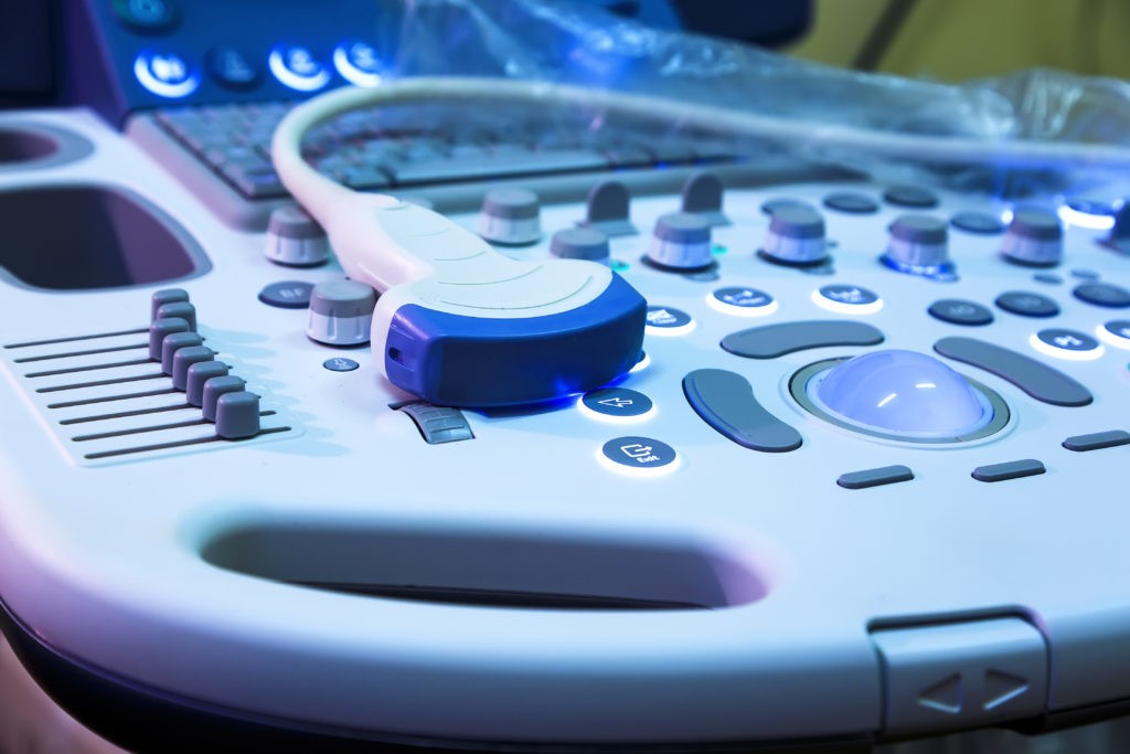
Regional anesthesia has enjoyed many advancements in its practice worldwide, but the most revolutionary advancement within the field has been the use of ultrasound.  Ultrasound has been shown to lead to more successful regional blocks over stimulation-based or paresthesia-based techniques1,2.  The use of ultrasound is able to provide safer regional anesthesia by helping providers avoid potential complications such as intravascular injection, nerve injury, or damage to local structures.   Since the adoption of ultrasound as the gold standard for regional anesthesia during the past few decades, performing nerve blocks has become significantly safer and increasingly useful for a wide variety of procedures.

A significant progression in ultrasound-guided regional anesthesia has been improved needle technology.  More echogenic needles can provide better visualization of the nerve with respect to the injection site, giving a greater chance for block success3.  This is accomplished by either creating dimples in the needle, forming a rough needle surface, or adjusting the polymeric makeup of the needle coating to reflect ultrasound beams better4.  These changes to the needle tip may work better for certain types of blocks, but they may prove disadvantageous for others.  For example, a needle reflective for a specific frequency range of ultrasound required for deep blocks may not be so advantageous when a different frequency is utilized for superficial blocks.  To this end, some needle manufacturers have introduced the ability to perform electromagnetic-guided needle-tip tracking in conjunction with ultrasound5.  While needle tip tracking has been found to improve provider confidence in an adequate block, this technology is very new and still has yet to gain widespread regulatory approval. 

In addition to improved needle quality for imaging, ultrasound technology itself has advanced far enough to significantly facilitate the ease and efficacy of regional anesthesia.  Over the past several years, transducer quality, improved image resolution, and better beam formation have all drastically increased the utility of ultrasound in providing accurate sonoanatomy for safe and effective blocks4.  In addition, ultrasound technology has ventured into newer modalities that may provide additional ease to performing regional anesthesia; three-dimensional ultrasound imaging can help elucidate anatomic structures easier, facilitate more precise needle movements, and map the spread of local anesthetic around the target area.

The use of ultrasound guidance of regional anesthesia has come a long way, and many more helpful advances are coming into widespread use.  Machine learning may play a key role in imaging processing, with nearly complete minimization of artifact and shadowing in order to provide better visualization of structures.  In addition, machine learning may eventually become advanced enough to automatically identify specific anatomic structures in real-time to aid the provider in reaching the intended target.  Through improvement of beam formation and image resolution to better penetrate structures with poor acoustic transmission, even neuraxial anesthesia can receive immense benefit from ultrasound guidance.  The use of automation may also be a key development in the advancement of regional anesthesia, helping providers maneuver the ultrasound probe or needle in order to administer the most precise block possible.  The practice of ultrasound-guided regional anesthesia has come so far since its first reported use in 19941; it is extremely likely that in another 25 years, the field will see immense improvements in patient safety, success rates, and utility for numerous clinical situations.

1.         Gray AT. Ultrasound-guided regional anesthesia: current state of the art. Anesthesiology. 2006;104(2):368-373, discussion 365A.

2.         Kapral S, Greher M, Huber G, et al. Ultrasonographic guidance improves the success rate of interscalene brachial plexus blockade. Reg Anesth Pain Med. 2008;33(3):253-258.

3.         Hebard S, Hocking G. Echogenic technology can improve needle visibility during ultrasound-guided regional anesthesia. Reg Anesth Pain Med. 2011;36(2):185-189.

4.         Henderson M, Dolan J. Challenges, solutions, and advances in ultrasound-guided regional anaesthesia. BJA Education. 2016;16(11):374-380.

5.         Kåsine T, Romundstad L, Rosseland LA, et al. Needle tip tracking for ultrasound-guided peripheral nerve block procedures-An observer blinded, randomised, controlled, crossover study on a phantom model. Acta Anaesthesiol Scand. 2019.

6.         Clendenen NJ, Robards CB, Clendenen SR. A standardized method for 4D ultrasound-guided peripheral nerve blockade and catheter placement. Biomed Res Int. 2014;2014:920538.

While it is possible to place catheters at various anatomic locations, they are not equivalent in terms of analgesic benefit. Likewise, analgesia during the infusion has been demonstrated in several high-quality studies, however positive effect on chronic postoperative pain is lacking in evidence. Interscalene and sciatic catheters provide very potent and satisfactory analgesia, whereas supraclavicular, axillary, and transversus abdominus plane catheters have not shown the same level of effect. Placing an infraclavicular catheter has shown promise from an analgesia standpoint, however the dose required often causes complete anesthesia and motor block of the limb. Femoral and lumbar plexus infusions have shown similar effect, causing weakness that may interfere with the recovery process. Studies have reported a fall risk as high as 5 times that of controls when continuous femoral block is placed.4,5 That is not to say these blocks should not be performed, however the patient should be counseled regarding the possible result of placing a PNC in these locations.

As with any invasive procedure, the risks and benefits must be considered, weighed, and disclosed to the patient. Due to the dependence of results of any procedure on provider technique, it is difficult make overarching statements regarding the incidence and prevalence, but severe complications are very rarely reported. Minor complications are reported at a rate similar to that of single-shot peripheral nerve blocks.2,4 Infusion failure has been reported anywhere from 0.5% to 26%, however it must again be noted that these studies exhibit significant heterogeneity in terms of technique, equipment, catheter location, and infusate regimen. If it difficult to generalize based on these limitations however it should be noted that the adoption of ultrasound guidance has significantly decreased the rate of reported block failure.

Nerve damage is a potential complication that may be anxiety provoking for the patient, however studying the true incidence of neuropathy attributable to use of regional anesthesia is difficult. This is partly due the fact that the surgery itself, tourniquet time, and the prolonged positioning without protective reflexes serve as risk factors for nerve damage. What is known from currently available evidence is that the rate of neuropathy is generally low and symptoms in most cases resolve within a year without treatment.5 Prior nerve injury in the planned location or history of neuropathy are relative contraindications due to the increased risk of recurrence or worsening of neuropathy.3 Professional athletes are not at increased risk, however it may be prudent to forego regional techniques given their dependence on reliable return of neuromuscular function and coordination for their occupation.

A relatively common complication of any perineural catheter is dislodgement or malpositioning leading to block failure, which often leads the provider to take extra measures to secure the catheter at the insertion point, and at the skin, either coiled, glued, anchored, or tunneled to prevent tension on the catheter being transmitted to the tip. There has also been a single report of leakage from the catheter site, causing contamination of the surgical field intraoperatively, but skin adhesives like 2-Octyl cyanoacrylate have been found to decrease the incidence of leakage.5 Extremely rare, however potentially fatal, is migration of the catheter into an intravascular, epidural, intrathecal, or intrapleural location.

Another potentially devastating complication of the use of an indwelling catheter of any type is infection, although the reported incidence is less than 1%. Patients at significant risk include those who are immunosuppressed or otherwise immunocompromised, male gender, diabetics, obese patients, victims of trauma, and those being treated in the ICU. Maintenance of a perineural catheter for more than 48hrs is a modifiable risk factor, while other factors such as aseptic drawing of medication, catheter tunneling, and use of aseptic dressings have not shown significance.Axillary, femoral, and even interscalene catheters have been reported to have higher risk of infection. Peri-catheter hematoma is also of concern, specifically in patients maintained on anticoagulants at the time of placement, manipulation, or removal of a neuraxial or “deep” perineural catheter, however there is conflicting evidence regarding this risk in most peripheral blocks. Some researchers have recommended substituting erector spinae, paravertebral, or transversus abdominis plane catheters for an epidural when concern for coagulopathy exists.

Finally, there are alternatives to a perineural catheter ranging from opioid-based analgesia, wound infiltration, cryoanalgesia, acupuncture, percutaneous nerve stimulation, single shot peripheral block with liposomal bupivacaine, or single shot neuraxial, each with its own unique set of risks and benefits.5 Careful selection of appropriate patients for peripheral nerve catheter has shown great success in minimizing the consumption of opioids, an emerging priority in healthcare policy as efforts are increased to eradicate the opioid epidemic. Moreover, research and innovation continue to expand the list of approved indications for placement of peripheral nerve catheter. There are still many questions which remain unanswered, heralding a need for on-going experimentation to expand our collective understanding of continuous regional anesthesia.

References

1. Ansbro FP. A method of continuous brachial plexus block. The American Journal of Surgery. 1946;71(6):716-722. https://www.sciencedirect.com/science/article/pii/000296104690219X. doi: 10.1016/0002-9610(46)90219-X.

2. Borgeat A, Ekatodramis G, Kalberer F, Benz C. Acute and nonacute complications associated with interscalene block and shoulder Surgery: A prospective study. Anesthes. 2001;95(4):875-880. https://anesthesiology.pubs.asahq.org/article.aspx?articleid=1944139. Accessed Dec 5, 2019.

3. Chang A, White BA. Peripheral nerve blocks. In: StatPearls. Treasure Island (FL): StatPearls Publishing; 2019. http://www.ncbi.nlm.nih.gov/books/NBK459210/. Accessed Dec 10, 2019.

4. Ilfeld B. Continuous peripheral nerve blocks: A review of the published evidence. Anesthesia & Analgesia. 2011;113(4):904-925. insights.ovid.com. Accessed Nov 25, 2019. doi: 10.1213/ANE.0b013e3182285e01.

5. Ilfeld B. Continuous peripheral nerve blocks: An update of the published evidence and comparison with novel, alternative analgesic modalities. Anesthesia & Analgesia. 2017;124(1):308-335. insights.ovid.com. Accessed Nov 25, 2019. doi: 10.1213/ANE.0000000000001581.

6. Joshi G, Gandhi K, Shah N, Gadsden JC, Corman SL. Peripheral nerve blocks in the management of postoperative pain: Challenges and opportunities. Journal of Clinical Anesthesia. 2016;35:524-529. http://www.ncbi.nlm.nih.gov/pubmed/27871587. Accessed Nov 25, 2019.

7. Luyet C, Seiler R, Herrmann G, Hatch GM, Ross S, Eichenberger U. Newly designed, self-coiling catheters for regional anesthesia-an imaging study. Reg Anesth Pain Med. 2011;36(2):171-176. https://rapm.bmj.com/content/36/2/171-176. Accessed Dec 12, 2019. doi: 10.1097/AAP.0b013e31820d431a.

8. Marhofer P, Greher M, Kapral S. Ultrasound guidance in regional anaesthesia. Br J Anaesth. 2005;94(1):7-17. https://academic.oup.com/bja/article/94/1/7/379332. Accessed Dec 2, 2019. doi: 10.1093/bja/aei002.

9. Nicolotti D, Iotti E, Fanelli G, Compagnone C. Perineural catheter infection: A systematic review of the literature. J Clin Anesth. 2016;35:123-128. Accessed Nov 25, 2019. doi: 10.1016/j.jclinane.2016.07.025.

10. Schnabel A, Meyer-Frießem CH, Zahn PK, Pogatzki-Zahn EM. Ultrasound compared with nerve stimulation guidance for peripheral nerve catheter placement: A meta-analysis of randomized controlled trials. British Journal of Anaesthesia. 2013;111(4):564-572. https://www.sciencedirect.com/science/article/pii/S0007091217323486. doi: 10.1093/bja/aet196. 11. Toledano RD, Tsen LC. Epidural catheter DesignHistory,innovations,and clinical implications. Anesthes. 2014;121(1):9-17. https://anesthesiology.pubs.asahq.org/article.aspx?articleid=1917668. Accessed Nov 25, 2019. doi: 10.1097/ALN.0000000000000239.

The technique for placement of nerve catheters has evolved over time, initially being done using anatomic landmarks and either subjective paresthesia, fascial “click” or fluoroscopic visualization. By the 1970s, the use of nerve stimulation was popularized and by 1978 the first use of ultrasound guidance for nerve block was reported by LaGrange et al.Unfortunately, the adequacy of analgesia obtained using either technique is operator and situation dependent. Whether the nerve is localized using a stimulating needle, stimulating catheter, or sonographically there remains the possibility of inadequate pain control in spite of adequate muscle stimulation or nerve visualization.

The location of the catheter tip is also of concern when placing and evaluating PNCs. The catheter tip may easily be mispositioned or become dislodged too far from the nerve to provide adequate effect postoperatively. However, if a large initial bolus was given through the catheter or via needle prior to advancing the catheter, mispositioning may not be noticed until the bolus clears and the infusion is started postoperatively. No studies have elucidated an optimal distance to thread the catheter past the tip, however increased coiling and knotting has been reported with catheters >5cm beyond the needle tip, thus the current recommendation is not to exceed 5cm distance beyond the needle.4 In 2011, there was a report of self-coiling catheters being developed to maintain close proximity of the catheter to the nerve, however these were not approved for human use.

After numerous studies comparing the effectiveness of nerve stimulation and ultrasound in placement of nerve catheters, meta-analysis was performed in 2013 demonstrating clear superiority for ultrasound guidance in terms of success rate and avoidance of vascular puncture. Time required to place a catheter and procedural pain were also found to be decreased.10

The local anesthetic most commonly infused through PNCs is either bupivacaine or ropivacaine, which are long-acting amide local anesthetics, however any of the available local anesthetics can be used, as long as an appropriate dosing regimen is followed. Opiates are also added frequently to the infusate to supplement a neuraxial blockade given the decreased likelihood of adverse side effects when the opioid Mu receptors are modulated directly at the level of the dorsal horn. Delivery regimens are based on a set bolus dose, continuous rate, or combination of the two. Bolus-only regimens will clearly decrease the total consumption of anesthetic, however there is evidence of superior analgesia when sciatic catheters are maintained on a basal infusion. Regarding the infusion system, electronic pumps are reported to be very consistent in terms of delivery of the selected basal rate (5% error), whereas elastomeric systems are found to over-infuse during the first 8 hours and the final hours of infusion.4 In addition, elastomeric devices cannot be refilled outside of a pharmacy, lack the option for bolus dosing, and lack any alarm mechanism. The major upside to an elastomeric pump is that is simplifies ambulatory dosing of a PNC in the postoperative period.

First reported in 1997, ambulatory continuous perineural catheters, have become much more common in recent years with the push to decrease healthcare cost and length of stay. Because local anesthetic is being administered in a non-monitored setting, care must be taken with patient selection to avoid those at high risk of poor drug clearance, infection, or misuse. It should be noted that young age is not a contraindication, as studies have found no increase in rate of complications among pediatric patients. Fall risk must also be considered as patients receiving ropivacaine via PNC have been found to have increased incidence of falls. There has also been report of PNC masking pain from a new metatarsal fracture after a fall. Patients receiving ambulatory infusions via perineural catheter should be followed closely, however there is currently no standard recommendation regarding a method or frequency.4 It is common practice to call patients daily while the catheter is in place to monitor analgesia, site appearance, remaining local anesthetic, and pump function. Patients are also given instructions on how to remove the catheter at home and are commonly instructed via telephone during the removal process.

References

1. Ansbro FP. A method of continuous brachial plexus block. The American Journal of Surgery. 1946;71(6):716-722. https://www.sciencedirect.com/science/article/pii/000296104690219X. doi: 10.1016/0002-9610(46)90219-X.

2. Borgeat A, Ekatodramis G, Kalberer F, Benz C. Acute and nonacute complications associated with interscalene block and shoulder Surgery: A prospective study. Anesthes. 2001;95(4):875-880. https://anesthesiology.pubs.asahq.org/article.aspx?articleid=1944139. Accessed Dec 5, 2019.

3. Chang A, White BA. Peripheral nerve blocks. In: StatPearls. Treasure Island (FL): StatPearls Publishing; 2019. http://www.ncbi.nlm.nih.gov/books/NBK459210/. Accessed Dec 10, 2019.

4. Ilfeld B. Continuous peripheral nerve blocks: A review of the published evidence. Anesthesia & Analgesia. 2011;113(4):904-925. insights.ovid.com. Accessed Nov 25, 2019. doi: 10.1213/ANE.0b013e3182285e01.

5. Ilfeld B. Continuous peripheral nerve blocks: An update of the published evidence and comparison with novel, alternative analgesic modalities. Anesthesia & Analgesia. 2017;124(1):308-335. insights.ovid.com. Accessed Nov 25, 2019. doi: 10.1213/ANE.0000000000001581.

6. Joshi G, Gandhi K, Shah N, Gadsden JC, Corman SL. Peripheral nerve blocks in the management of postoperative pain: Challenges and opportunities. Journal of Clinical Anesthesia. 2016;35:524-529. http://www.ncbi.nlm.nih.gov/pubmed/27871587. Accessed Nov 25, 2019.

7. Luyet C, Seiler R, Herrmann G, Hatch GM, Ross S, Eichenberger U. Newly designed, self-coiling catheters for regional anesthesia-an imaging study. Reg Anesth Pain Med. 2011;36(2):171-176. https://rapm.bmj.com/content/36/2/171-176. Accessed Dec 12, 2019. doi: 10.1097/AAP.0b013e31820d431a.

8. Marhofer P, Greher M, Kapral S. Ultrasound guidance in regional anaesthesia. Br J Anaesth. 2005;94(1):7-17. https://academic.oup.com/bja/article/94/1/7/379332. Accessed Dec 2, 2019. doi: 10.1093/bja/aei002.

9. Nicolotti D, Iotti E, Fanelli G, Compagnone C. Perineural catheter infection: A systematic review of the literature. J Clin Anesth. 2016;35:123-128. Accessed Nov 25, 2019. doi: 10.1016/j.jclinane.2016.07.025.

10. Schnabel A, Meyer-Frießem CH, Zahn PK, Pogatzki-Zahn EM. Ultrasound compared with nerve stimulation guidance for peripheral nerve catheter placement: A meta-analysis of randomized controlled trials. British Journal of Anaesthesia. 2013;111(4):564-572. https://www.sciencedirect.com/science/article/pii/S0007091217323486. doi: 10.1093/bja/aet196.

11. Toledano RD, Tsen LC. Epidural catheter DesignHistory,innovations,and clinical implications. Anesthes. 2014;121(1):9-17. https://anesthesiology.pubs.asahq.org/article.aspx?articleid=1917668. Accessed Nov 25, 2019. doi: 10.1097/ALN.0000000000000239.

A continuous peripheral nerve block (CPNB) or peripheral nerve catheter (PNC) is essentially an indwelling tubular device inserted via a needle through the skin, with orifices at each end for injection and delivery of local anesthetic around nerve or within a fascial plane known to contain traversing neural elements. Since its development, there has been a multitude of iterations and modifications, creating what is now an essential device in the prevention and treatment of postoperative pain after major surgery. The use of catheters for the infusion of local anesthetic extends back to 1931, when Dr. Eugen Aubrel, a Romanian professor of obstetrics and gynecology, used a silk ureteral catheter to deliver a continuous lumboaortic plexus block for the first stage of labor. Subsequently, the indwelling Lemmon needle was developed by Dr. William Lemmon and later modified by Drs. Robert Hingson and Waldo Edwards to provide a method of delivering continuous caudal anesthesia via a malleable stainless-steel needle attached to a syringe via long tubing.

After reports of the limitations associated with the modified Lemmon needle were published, including migration and breakage of the needle, Dr. Edward Tuohy applied the ureteral catheter to continuous spinal anesthesia in the 1940s. At the time, the catheter was delivered via a 15-gauge Barker needle, which upon removal was sequentially sterilized but not reused more than ten times per Dr. Tuohy’s recommendation. He subsequently developed the Tuohy needle, which is still in use today. Nylon catheters later replaced their silk predecessors after a case of meningitis was reported by Dr. Samuel Manalan in that same decade.

Several modifications to needle size, type, and technique were made over the following years until Dr. Manuel Curbelo, a Cuban anesthesiologist who had visited the Mayo Clinic three years prior, described an adaptation of the Tuohy needle to achieve peridural passage of an indwelling catheter in 1949 to provide anesthesia for surgeries below the neck. Drs. Charles Flowers, Louis Hellman, and Robert Hingson collaborated later that year to describe the use of a continuous epidural catheter, threaded through a 16-gauge Tuohy needle into the second lumbar interspace, for vaginal and cesarean deliveries, another technique which remains in use today, with minor modifications. The continuous peripheral nerve block was first described in 1946 by F. Paul Ansbro for upper extremity surgeries1. By 1951, Sarnoff and Sarnoff reported using a continuous nerve block to treat intractable hiccups, demonstrating the diversity of experimentation with regional anesthesia during that fruitful period in the history of anesthesia.4

Regarding the current practice in management of perineural catheters, there are several considerations and potential complications which must be accounted for in addition to the standard principles used to guide management of all nerve blocks. The indications for use of PNCs are essentially the same regarding surgical anesthesia, with the addition that the catheter allows for continuous delivery of dilute local anesthetic for extended postoperative analgesia. This is useful for cases where postoperative pain is expected to be persistently high and will be inadequately controlled on intravenous and/or oral analgesics. PNCs are also useful in situations where standard dosing of opioid analgesics is contraindicated or poorly tolerated (e.g. opioid tolerant or active substance abuse, severe PONV, severe OSA). In addition, continuous regional techniques have been used to treat vasospasm (Raynaud’s phenomenon, digital injury/reimplantation), CRPS, phantom limb pain, terminal cancer pain, and trigeminal neuralgia. While there are several useful clinical applications for CPNB, the most rigorously tested of these is for perioperative pain control. There remains a paucity of level-I studies evaluating the effectiveness of PNCs in these other applications, however there are several trials underway to this effect. Given the increased risk for potential complications from an indwelling device, PNCs are typically preserved for cases when single-shot nerve blocks along with other non-invasive analgesics will be inadequate and the benefit of placing a catheter outweighs the potential risks. Absolute contraindications to placement of a PNC are few and include patient refusal, allergy to local anesthetic, or inability to cooperate with procedure. Relative contraindications include active infection or preexisting nerve deficit in the intended site or distribution, and coagulopathy, especially when the planned block is in a non-compressible location.

References

1. Ansbro FP. A method of continuous brachial plexus block. The American Journal of Surgery. 1946;71(6):716-722. https://www.sciencedirect.com/science/article/pii/000296104690219X. doi: 10.1016/0002-9610(46)90219-X.

2. Borgeat A, Ekatodramis G, Kalberer F, Benz C. Acute and nonacute complications associated with interscalene block and shoulder Surgery: A prospective study. Anesthes. 2001;95(4):875-880. https://anesthesiology.pubs.asahq.org/article.aspx?articleid=1944139. Accessed Dec 5, 2019.

3. Chang A, White BA. Peripheral nerve blocks. In: StatPearls. Treasure Island (FL): StatPearls Publishing; 2019. http://www.ncbi.nlm.nih.gov/books/NBK459210/. Accessed Dec 10, 2019.

4. Ilfeld B. Continuous peripheral nerve blocks: A review of the published evidence. Anesthesia & Analgesia. 2011;113(4):904-925. insights.ovid.com. Accessed Nov 25, 2019. doi: 10.1213/ANE.0b013e3182285e01.

5. Ilfeld B. Continuous peripheral nerve blocks: An update of the published evidence and comparison with novel, alternative analgesic modalities. Anesthesia & Analgesia. 2017;124(1):308-335. insights.ovid.com. Accessed Nov 25, 2019. doi: 10.1213/ANE.0000000000001581.

6. Joshi G, Gandhi K, Shah N, Gadsden JC, Corman SL. Peripheral nerve blocks in the management of postoperative pain: Challenges and opportunities. Journal of Clinical Anesthesia. 2016;35:524-529. http://www.ncbi.nlm.nih.gov/pubmed/27871587. Accessed Nov 25, 2019.

7. Luyet C, Seiler R, Herrmann G, Hatch GM, Ross S, Eichenberger U. Newly designed, self-coiling catheters for regional anesthesia-an imaging study. Reg Anesth Pain Med. 2011;36(2):171-176. https://rapm.bmj.com/content/36/2/171-176. Accessed Dec 12, 2019. doi: 10.1097/AAP.0b013e31820d431a.

8. Marhofer P, Greher M, Kapral S. Ultrasound guidance in regional anaesthesia. Br J Anaesth. 2005;94(1):7-17. https://academic.oup.com/bja/article/94/1/7/379332. Accessed Dec 2, 2019. doi: 10.1093/bja/aei002.

9. Nicolotti D, Iotti E, Fanelli G, Compagnone C. Perineural catheter infection: A systematic review of the literature. J Clin Anesth. 2016;35:123-128. Accessed Nov 25, 2019. doi: 10.1016/j.jclinane.2016.07.025.

10. Schnabel A, Meyer-Frießem CH, Zahn PK, Pogatzki-Zahn EM. Ultrasound compared with nerve stimulation guidance for peripheral nerve catheter placement: A meta-analysis of randomized controlled trials. British Journal of Anaesthesia. 2013;111(4):564-572. https://www.sciencedirect.com/science/article/pii/S0007091217323486. doi: 10.1093/bja/aet196. 11. Toledano RD, Tsen LC. Epidural catheter DesignHistory,innovations,and clinical implications. Anesthes. 2014;121(1):9-17. https://anesthesiology.pubs.asahq.org/article.aspx?articleid=1917668. Accessed Nov 25, 2019. doi: 10.1097/ALN.0000000000000239.

Cardiovascular disease, which is the leading cause of death for both men and women in the United States,1 refers to various types of heart conditions that affect health and mortality. These conditions include coronary artery disease, cerebrovascular disease (stroke), congenital cardiovascular defects, heart rhythms, sudden cardiac arrest, cardiomyopathy, heart failure and more.2 According to the Centers for Disease Control and Prevention (CDC), having an unhealthy diet, sedentary lifestyle, high cholesterol, high blood pressure or diabetes can increase one’s risk of heart disease, as can smoking tobacco.3 Additionally, 0.4 to 1 per 100 United States infants are born with congenital heart disease, ranging from mild asymptomatic lesions to fatal conditions.2 Given the ubiquity of heart disease, medical providers must consider the complexity of a patient with cardiovascular issues. Anesthesiology practitioners, for example, need to account for cardiovascular disease before, during and after surgery to avoid complications or even mortality. 

Risks associated with anesthesia for patients with cardiovascular disease are common in children as well as adults. Ramamoorthy et al. used data from the Pediatric Perioperative Cardiac Arrest Registry to analyze 373 anesthesia-related cardiac arrests in children, 34 percent of whom had congenital or acquired heart disease.4 The authors found that children with heart disease were sicker than those without heart disease at the time of cardiac arrest and had a higher mortality rate.4 Though most of these events occurred during surgery, another study found that children with congenital heart disease could go into cardiac arrest throughout the perioperative period.5 In their paper, Cannesson et al. stress the risk of stroke, thrombosis (blood clots), heart failure and dysrhythmia during surgery for adults with congenital heart disease,6 while Odegard et al. emphasize cardiac arrest in similar patients.7 For children and adults with heart disease, the risks of cardiovascular or cerebrovascular complications in surgery are high. 

Given these risks, the anesthesiologist should adequately prepare the patient for surgery. Because preoperative fasting may increase risk of cerebrovascular thrombosis, the anesthesia provider should preoperatively assess the patient’s coagulation system and consistently hydrate the patient with intravenous fluids.6 The anesthesiologist may also have to premedicate the patient with anxiolytics and hypnotics in order to prepare the cardiovascular patient for a stressful surgery.6 For patients with congenital heart defects, the anesthesiologist must become familiar with the patient’s unique physiology in order to adequately prepare for a procedure.8 Some hospitals may apply strict guidelines for care or use technology, such as computer algorithms, to assess anesthesia-related risks in children with congenital heart disease.5 Preoperative preparation of patients with cardiovascular disease, such as adequate hydration and analysis of the patient’s particular condition, are necessary roles of the anesthesia provider. 

During surgery, the anesthesia practitioner is responsible for collaborating with other health professionals to ensure a seamless procedure.8 This includes vigilantly monitoring the patient to maintain steady blood pressure throughout and after the procedure.9 In children with congenital heart disease, anesthesia-related cardiac arrests occur most commonly during the procedure (as opposed to before or after), so anesthesia providers should constantly be aware of a patient’s vital signs.4 Also, using the right type of medication and anesthesia for a specific cardiovascular disease is essential to preventing complications or mortality. Studies suggest using diazepam,10 midazolam11 and/or a combination of epidural anesthesia and light general anesthesia12 to provide rapid, stable induction of anesthesia in patients with heart disease. Meanwhile, Russell et al. found that sevoflurane may have advantages over halothane in maintaining hemodynamic stability for children with congenital heart disease.13 

Risk of complications during surgery are high for patients with congenital or acquired heart disease. Given the possibility of anesthesia-related cardiac arrest or stroke, anesthesia providers must assess and monitor patients throughout the perioperative period. Before a procedure, the anesthesia practitioner should evaluate the patient’s anatomy and particularities of the heart disease, and provide treatments such as hydration and premedication. During surgery, provision of an appropriate anesthetic agent and collaboration with other professionals in the procedure room are important duties of the anesthesiologist. Furthermore, consistent vital signs monitoring is crucial during and after surgery. 

1. Centers for Disease Control and Prevention. Heart Disease in the United States. Heart Disease Facts 2019; https://www.cdc.gov/heartdisease/facts.htm

2. Benjamin EJ, Virani SS, Callaway CW, et al. Heart Disease and Stroke Statistics—2018 Update: A Report From the American Heart Association. Circulation. 2018;137(12):e67–e492. 

3. National Center for Chronic Disease Prevention and Health Promotion. Know the Facts About Heart Disease. Atlanta, GA: Centers for Disease Control and Prevention;2019. 

4. Ramamoorthy C, Haberkern CM, Bhananker SM, et al. Anesthesia-Related Cardiac Arrest in Children with Heart Disease: Data from the Pediatric Perioperative Cardiac Arrest (POCA) Registry. Anesthesia & Analgesia. 2010;110(5):1376–1382. 

5. Taylor D, Habre W. Risk associated with anesthesia for noncardiac surgery in children with congenital heart disease. Pediatric Anesthesia. 2019;29(5):426–434. 

6. Cannesson M, M.D., Earing Michael G, M.D., Collange V, M.D., Kersten Judy R, M.D., F.A.C.C. Anesthesia for Noncardiac Surgery in Adults with Congenital Heart Disease. Anesthesiology: The Journal of the American Society of Anesthesiologists. 2009;111(2):432–440. 

7. Odegard KC, DiNardo JA, Kussman BD, et al. The Frequency of Anesthesia-Related Cardiac Arrests in Patients with Congenital Heart Disease Undergoing Cardiac Surgery. Anesthesia & Analgesia. 2007;105(2):335–343. 

8. Gottlieb EA, Andropoulos DB. Anesthesia for the patient with congenital heart disease presenting for noncardiac surgery. Current Opinion in Anesthesiology. 2013;26(3):318–326. 

9. Howell SJ, Sear JW, Foëx P. Hypertension, hypertensive heart disease and perioperative cardiac risk. BJA: British Journal of Anaesthesia. 2004;92(4):570–583. 

10. Samuelson PN, Reves JG, Kouchoukos NT, Smith LR, Dole KM. Hemodynamic responses to anesthetic induction with midazolam or diazepam in patients with ischemic heart disease. Anesthesia and Analgesia. 1981;60(11):802–809. 

11. Middlehurst RJ, Gibbs A, Walton G. Cardiovascular risk: The safety of local anesthesia, vasoconstrictors, and sedation in heart disease. Anesthesia Progress. 1999;46(4):118–123. 

12. Reiz S, Bålfors E, Sørensen MB, Häggmark S, Nyhman H. Coronary Hemodynamic Effects of General Anesthesia and Surgery: Modification by Epidural Analgesia in Patients with Ischemic Heart Disease. Regional Anesthesia: The Journal of Neural Blockade in Obstetrics, Surgery, & Pain Control. 1982;7(Suppl 4):S8–S18. 

13. Russell IA, Miller Hance WC, Gregory G, et al. The Safety and Efficacy of Sevoflurane Anesthesia in Infants and Children with Congenital Heart Disease. Anesthesia & Analgesia. 2001;92(5):1152–1158. 

Obesity, defined as a body mass index (BMI) at or above 30 kg/m2, has become an “epidemic” according to contemporary researchers.1 The Centers for Disease Control and Prevention (CDC) recognized the obesity epidemic as a national problem in 1999, when it published a series of maps showing rapid changes in the prevalence of obesity.2 Even before obesity began costing the United States over $117 billion per year in medical costs,1 medical providers had been aware of the increased medical risks for obese patients. In fact, research as early as the 1960s acknowledged the effects of obesity on complications following general anesthesia.3 The worldwide increase in obesity has required anesthesiology practitioners to take further considerations while their patients undergo various procedures.

For one, obesity can affect accuracy of anesthesia dosing. Because of large differences between lean body weight (LBW) and total body weight (TBW) in obese patients,4,5 the pharmacokinetics (i.e., the way drugs are absorbed by the body) and pharmacodynamics (i.e., the effects of drugs and their mechanisms of action) of anesthetic drugs are altered.5 For example, several researchers have debated whether dosage adjustments in obese patients for Propofol, a common injectable general anesthetic, should be based on LBW or TBW.4-6 For many other anesthesia drugs, such as opioids7 and inhaled anesthetics,6 best practices for obese patients remain unclear. Overall, the complexity of obese patients’ altered lipid levels can affect dosing of anesthetic drugs in unpredictable ways.

Additionally, obese patients often show comorbidity, or the simultaneous presence of more than one chronic medical condition. Disorders that are comorbid with obesity, such as obstructive sleep apnea (OSA),8 can cause poor outcomes in anesthesiology. Some studies show that OSA and obesity may even share genetic risk factors,9,10 which may make OSA common in obesity and thus affect anesthesia for obese patients. Given that OSA is marked by upper airway blockages, brief periods of breathing cessation and lack of oxygenation,11 it can have harmful effects on anesthesia administration. In obese patients, who have more fatty tissue in the larynx, poor respiratory outcomes during anesthesia include intubation failure and respiratory obstruction soon after extubation.12 Also, the provision of opioids during procedures can add further respiratory and arousal depression, resulting in more issues for patients who already face obesity and OSA.12 Given the respiratory problems associated with OSA and obesity, anesthesiologists must keep in mind positioning of obese patients, using regional anesthesia, monitoring vital signs vigilantly and minimizing oxygen loss during anesthesia.

OSA and pharmacological changes only represent two of the main issues faced by obese patients undergoing anesthesia. Other challenges the anesthesiologist must confront include issues with cardiac, respiratory and metabolic systems and perioperative management (before and after surgery).14 Many studies also address complications facing certain populations with obesity and specific situations in which anesthesia may be necessary. For example, patients with obesity have increased complications during pregnancy and childbirth, a higher rate of caesarean sections and potential for difficulties with emergency anesthesia.15 Thus, the data suggest that epidural anesthesia during labor be well-planned and administered early and often.15-17 Other research has focused on anesthesia management in obese children, which involves careful preanesthesia assessment, changes in drug selection and consideration of comorbidities.18-20 Further studies have addressed the potential advantages and disadvantages of using regional instead of general anesthesia in obese patients when appropriate,6,21 as well as the effects obesity may have on regional anesthesia techniques.6,21,22 Evidently, anesthetic management of patients with obesity may vary based on patients’ ages, stages of gestation and types of anesthesia.

The obesity epidemic has effects on the pharmacology and risks of anesthesia administration. Anesthesia providers who care for patients with obesity must account for altered body weight, comorbidities that affect airway function and factors such as pregnancy, childhood and anesthesia location. As obesity affects more patients worldwide, anesthesiology practitioners must take more precautions before, during and after administering anesthetic drugs.

1.         Stein CJ, Colditz GA. The Epidemic of Obesity. The Journal of Clinical Endocrinology & Metabolism. 2004;89(6):2522–2525.

2.         Dietz WH. The Response of the US Centers for Disease Control and Prevention to the Obesity Epidemic. Annual Review of Public Health. 2015;36(1):575–596.

3.         Gould AB, Jr. Effect of obesity on respiratory complications following general anesthesia. Anesthesia and Analgesia. 1962;41:448–452.

4.         Casati A, Putzu M. Anesthesia in the obese patient: Pharmacokinetic considerations. Journal of Clinical Anesthesia. 2005;17(2):134–145.

5.         Dong D, Peng X, Liu J, Qian H, Li J, Wu B. Morbid Obesity Alters Both Pharmacokinetics and Pharmacodynamics of Propofol: Dosing Recommendation for Anesthesia Induction. Drug Metabolism and Disposition. 2016;44(10):1579–1583.

6.         Ingrande J, Lemmens HJM. Anesthetic Pharmacology and the Morbidly Obese Patient. Current Anesthesiology Reports. 2013;3(1):10–17.

7.         Egan TD, MD, Huizinga B, MD, Gupta SK, PhD, et al. Remifentanil Pharmacokinetics in Obese versus Lean Patients. Anesthesiology: The Journal of the American Society of Anesthesiologists. 1998;89(3):562–573.

8.         Wittels EH, Thompson S. Obstructive sleep apnea and obesity. Otolaryngologic Clinics of North America. 1990;23(4):751–760.

9.         Patel SR. Shared genetic risk factors for obstructive sleep apnea and obesity. Journal of Applied Physiology. 2005;99(4):1600–1606.

10.       Palmer LJ, Buxbaum SG, Larkin EK, et al. Whole Genome Scan for Obstructive Sleep Apnea and Obesity in African-American Families. American Journal of Respiratory and Critical Care Medicine. 2004;169(12):1314–1321.

11.       Alkhalil M, Schulman E, Getsy J. Obstructive sleep apnea syndrome and asthma: What are the links? Journal of Clinical Sleep Medicine. 2009;5(1):71–78.

12.       Benumof JL. Obesity, sleep apnea, the airway and anesthesia. Current Opinion in Anesthesiology. 2004;17(1):21–30.

13.       Passannante AN, Rock P. Anesthetic Management of Patients with Obesity and Sleep Apnea. Anesthesiology Clinics of North America. 2005;23(3):479–491.

14.       Domi R, Laho H. Anesthetic challenges in the obese patient. Journal of Anesthesia. 2012;26(5):758–765.

15.       Eskandr A, Mostafa A, Metwally A, Afify N. Challenge of morbid obesity in obstetric anesthesia. Menoufia Medical Journal. 2015;28(2):308–314.

16.       Wallace DH, Santos R, Currie JM, Gilstrap LC. Indirect Sonographic Guidance for Epidural Anesthesia in Obese Pregnant Patients. Regional Anesthesia: The Journal of Neural Blockade in Obstetrics, Surgery, & Pain Control. 1992;17(4):233–236.

17.       Vallejo MC. Anesthetic management of the morbidly obese parturient. Current Opinion in Anesthesiology. 2007;20(3):175–180.

18.       Chidambaran V, Tewari A, Mahmoud M. Anesthetic and pharmacologic considerations in perioperative care of obese children. Journal of Clinical Anesthesia. 2018;45:39–50.

19.       Setzer N, Saade E. Childhood obesity and anesthetic morbidity. Pediatric Anesthesia. 2007;17(4):321–326.

20.       Baker S, Yagiela JA. Obesity: A complicating factor for sedation in children. Pediatric Dentistry. 2006;28(6):487–493.

21.       Nielsen Karen C, M.D., Guller U, M.D., M.H.S., Steele Susan M, M.D., Klein Stephen M, M.D., Greengrass Roy A, M.D., F.R.C.P., Pietrobon R, M.D., Ph.D. Influence of Obesity on Surgical Regional Anesthesia in the Ambulatory Setting: An Analysis of 9,038 Blocks. Anesthesiology: The Journal of the American Society of Anesthesiologists. 2005;102(1):181–187.

22.       Parra MC, Loftus RW. Obesity and Regional Anesthesia. International Anesthesiology Clinics. 2013;51(3):90–112.

Introduced in early July 2019 to Congress, H.R. bill 3630—known as the No Surprises Act—is intended to limit out-of-network rates that may be charged to individuals insured by healthcare plans for certain emergency and non-emergency services [1]. The bipartisan No Surprises Act (shorthand for ‘preventing surprise medical bills’) was spearheaded by Energy and Commerce Chairman Frank Pallone, Jr. (D-NJ) and Ranking Member Greg Walden (R-OR). The bill saw full passage in the Energy and Commerce Committee on July 17, 2019 and was then integrated into H.R. 2328, the Reauthorizing and Extending America’s Community Health Act [2, 3]. The impetus for the legislation arose from cases in which plan-holding patients have been surprised with unanticipated medical charges after receiving emergency services. Despite the fact that services are provided at in-network hospitals, patients may still receive care from out-of-network medical professionals—most commonly, unknowingly

Firstly, the proposed legislation addresses this issue by requiring that healthcare coverage plans covering emergency services bill members no more than the median in-network rate for emergency services, regardless of provider network status. Secondly, the bill goes further by prohibiting medical insurers from charging plan holders more than the average in-network cost for non-emergency services administered by out-of-network providers at in-network facilities [1, 3, 4]. In short, under the No Surprises Act, out-of-network providers are barred from billing patients the difference between in-network and out-of-network rates for emergency services.

Additionally, for non-emergency services, the bill protects plan holders from being charged for the difference in rates for non-emergency medical services rendered at in-network facilities, unless a patient is provided with specific notice and written-consent requirements to ensure that they are aware of the possibility of incurring out-of-network fees prior to receiving treatment [1, 2, 4]. However, an important caveat regarding the billing of non-emergency services in special cases is built into the legislation. This is that out-of-network providers may not bill plan holders for the difference between the rates for out-of-network and in-network non-emergency services if the provider whose services are required is the only professional available and/or qualified to deliver necessary services or treatments. Still, the provider in question must be based at an in-network facility for the proposed rate-capping to apply 

As part of the No Surprises Act, the Department of Health and Human Services is required to disburse grants to create and manage All Payer Claims Databases intended to make insurance claims and payment data public. Additionally, the bill calls for healthcare insurers to publish provider directories and instructs the Government Accountability Office and the Department of Labor to research and disclose information pertinent to commercial healthcare markets [1, 2, 4]. With similar aims as those encompassed by the No Surprises Act, the Senate Health, Education, Labor and Pensions (HELP) Committee passed the Lower Health Care Costs Act (S. 1895). Moving forward, leaders of the House Energy and Commerce Committee and Senate Health, Education, Labor and Pensions (HELP) Committee are in conversation over compatible and shared goals in their approaches [2, 3]. Such collaborations are of utmost importance as there exists significant potential for cost savings; for the No Surprises Act, an estimated $21.9 billion in savings is projected over 10 years [4, 5]. With impending expiration of health provisions and considerable monetary implications at stake, legislation intended to reduce surprise billing is likely to garner attention as the year closes 

Pallone, and Frank. “H.R.3630 – 116th Congress (2019-2020): No Surprises Act.” Congress.gov, July 11, 2019. https://www.congress.gov/bill/116th-congress/house-bill/3630.

[2] “House Energy and Commerce Committee Advances Surprise Billing Legislation.” AANS, n.d. https://www.aans.org/AANS-E-News/2019/8-30-E-news/House-Energy-and-Commerce-Committee-Advances-Surprise-Billing-Legislation.

[3] “Pallone & Walden on Committee Passage of No Surprises Act.” Democrats, Energy and Commerce Committee, July 17, 2019. https://energycommerce.house.gov/newsroom/press-releases/pallone-walden-on-committee-passage-of-no-surprises-act.

[4] “H.R. 3630: No Surprises Act.” GovTrack.us, July 9, 2019. https://www.govtrack.us/congress/bills/116/hr3630/text.

[5] “What’s New in Washington – October 2019 – Strategy – United States.” What’s New in Washington – October 2019 – Strategy – United States, October 24, 2019. http://www.mondaq.com/unitedstates/x/856814/Industry Updates Analysis/What’s New in Washington October 2019.[6] Keisling, Jonathan. “Assessing the Legislative Responses to Surprise Billing and Other Transparency Issues.” AAF, July 10, 2019. https://www.americanactionforum.org/insight/assessing-the-legislative-responses-to-surprise-billing-and-other-transparency-issues/.

Ventilation strategies for infants and children are also important to tailor to each patient as there is greater risk for barotrauma and atelectrauma. The closing capacity of an infant’s lungs may be higher than functional residual capacity, making them prone to atelectasis and desaturation. Ventilation should be monitored continuously using end tidal CO2 monitoring intraoperatively as well as chest-rise and auscultation in the perioperative phases. In children at increased risk of apnea, a portable oxygen saturation monitor can be particularly useful during transport between care areas.

Induction is a very critical phase which can be approached in various ways, but may be dictated by the patient’s age, anxiety level, aspiration risk, or airway anatomy. Infants, toddlers, young children, and adolescents each require slightly different approaches to induction, depending on presence of peripheral access, level of anxiety, willingness to cooperate with mask induction, and reaction to the smell of the mask and sevoflurane. In some instances, nitrous oxide is used to “stun” the patient prior to use of sevoflurane to prevent the need for forceful restraint on induction. In general, children have a higher volume of distribution and thus require increased doses of intravenous anesthetic to achieve induction of anesthesia (the same is true for neuromuscular blockade). Inhalational induction is commonly used unless there is a concerning component to the patient’s status such as a difficult airway or full stomach, in which case rapid sequence intubation is indicated and a pre-induction IV must be placed. Premedication with midazolam or parental presence can be very effective in preventing an uncontrolled induction that may require forceful restraint of the child’s limbs and head.2 Such an event can be particularly harmful in patients with a preexisting injury or congenital malformation. The audiovisual environment of the operating room at time of induction must also be managed. Parents, if available, should be readily visible to the child, the OR should be quiet, and nursing staff should not be busy moving in the background. All available staff should be attentive and prepared to assist if needed during this critical phase. A pre-induction checklist is useful to prevent the need to search or ask for equipment during induction. Young children are at increased risk of laryngospasm if depth of anesthesia is lost due to forceful or interrupted mask induction.5 Thus, succinylcholine 3mg/kg with an IM needle should be readily available and the provider should already have the dose required in mind prior to induction. The use of succinylcholine must take into account the possibility of an undiagnosed muscular dystrophy in infants, which will present as hyperkalemia, rhabdomyolysis, and possibly renal dysfunction perioperatively. Finally, inhaled induction poses a risk of hypotension if large doses of volatile anesthetic are rapidly delivered to the point of causing bradycardia. This is due to the pediatric heart’s lack of dynamic compliance and its dependence on preload and heart rate for maintenance of cardiac output.

Maintenance of anesthesia in children has its own unique set of considerations. Again, there is a higher volume of distribution, shorter half-life, and quicker clearance for many of the commonly used IV anesthetics; thus their weight-based dose requirements are often higher. The minimum alveolar concentration (MAC) required for general anesthesia is also higher in children, peaking in infants up to around six months old and decreasing thereafter. Propofol, for example, requires administration of twice the adult infusion rate for maintenance of a total intravenous anesthetic and at least 50% increase in bolus doses for induction. Regarding neuromuscular blockade, neonates are actually more sensitive to paralysis and while the required induction dose may be relatively higher, the maintenance dose should be decreased. Children have higher incidence of postoperative nausea and vomiting (PONV) as well as emergence delirium; this must be considered and planned for preoperatively in order to prevent postoperative complications. In fact, children who experience preoperative anxiety are 3.5 times more likely to exhibit negative behavior postoperatively.2 Medications such as ondansetron and dexmedetomidine are useful for prophylaxis of PONV and emergence delirium respectively but must be dosed and timed appropriately for optimum effect. Temperature management must also be conducted aggressively It begins preoperatively with warming of the entire OR suite, and includes the use of forced air warmers and under-body blankets placed appropriately to avoid thermal injury. Pediatric patients are more prone to heat loss in the perioperative period due to their relatively larger head and higher surface area to mass ratio but are also more vulnerable to skin burns if warming devices are placed too close to the skin.

Emergence and extubation are just as critical as induction and intubation, given a child’s predisposition to PRAEs. The primary goal during this phase is to ensure adequate spontaneous oxygenation and ventilation, avoid laryngospasm, and to remove the airway either before or after Stage II, as this excitatory period is when children are most prone to PRAEs.3 Choosing between awake and deep extubation can be challenging in complex cases but is typically guided by patient factors such as: type of surgery, presence of difficult/airway, risk of aspiration or obstruction, risk of hematoma or damage to surgical closure with coughing, and risk of bronchospasm. For surgeries about the airway and neck, deep extubation is preferred to avoid hematoma formation which itself can completely obstruct the pediatric airway. If the airway is tenuous for any reason, awake extubation is preferred as this minimizes the potential for obstruction during emergence. Prior to deep extubation, adequate spontaneous ventilation, reversal of paralysis, and depth of anesthesia >1 MAC must be confirmed. Oropharyngeal and/or orogastric suctioning and manipulation of the airway within the trachea are commonly performed to remove any potentially stimulating secretions and assess patient reaction to mucosal stimulation. For awake extubation, confirmation of adequate spontaneous ventilation, reversal of paralysis, and observation for conjugate gaze and grimacing with suctioning should be performed to confirm recovery from anesthesia. Should laryngospasm occur in either setting, delivery of 100% oxygen with positive pressure ventilation via a mask with a tight seal will break mild laryngospasm; however more severe episodes may require increasing the depth of anesthesia with propofol or paralysis with succinylcholine. Thereafter, the patient should be supported with mask ventilation and/or intubation.

In planning the setting of surgery and postoperative care for children, the risk of postoperative apnea must be considered. Those patients with a history of obstructive sleep apnea or prematurity, and infants less than <44 weeks post-menstrual age are at increased risk of this complication and should be strongly considered for postoperative admission.4,8

In summary, pediatric anesthesiology is a challenging yet rewarding field that incorporates much of what providers have learned from their adult patients, but also requires the addition of several special considerations and a thorough understanding of pediatric anatomy, physiology, and pharmacokinetics. Some of the challenges seen in caring for adult patients, such as preoperative anxiety and adverse respiratory events, are more commonly seen in children and must be anticipated. Every aspect of the anesthesiologist’s approach to a case, from preoperative evaluation to extubation and recovery must be modified to the unique needs and constraints presented by children as they traverse the stages of their development. Success or failure depends heavily on how effective one is at recognizing and either preventing or diagnosing and treating these common anesthetic challenges and complications.

1. Apfelbaum, Caplan, Connis, Epstein, Nickinovich, Warner. Practice guidelines for preoperative fasting and the use of pharmacologic agents to reduce the risk of pulmonary aspiration: Application to healthy patients undergoing elective procedures: An updated report by the american society of anesthesiologists task force on preoperative fasting and the use of pharmacologic agents to reduce the risk of pulmonary aspiration. Anesthesiology. 2011;114(3):495-511. doi: 10.1097/ALN.0b013e3181fcbfd9.

2. Banchs RJ, MD, Lerman, Jerrold, MD, FRCPC, FANZCA. Preoperative anxiety management, emergence delirium, and postoperative behavior. Anesthesiology Clinics. 2014;32(1):1-23. doi: 10.1016/j.anclin.2013.10.011.

3. Butz SF. Pediatric ambulatory anesthesia challenges. Anesthesiol Clin. 2019;37(2):289-300. Accessed Nov 3, 2019. doi: 10.1016/j.anclin.2019.01.002.

4. Coté CJ, Kelly DH. Postoperative apnea in a full-term infant with a demonstrable respiratory pattern abnormality. Anesthesiology. 1990;72(3):559-561. Accessed Nov 4, 2019. doi: 10.1097/00000542-199003000-00027.

5. De Francisci G, Papasidero AE, Spinazzola G, et al. Update on complications in pediatric anesthesia. Pediatric reports. 2013;5(1):e2. doi: 10.4081/pr.2013.e2.

6. Fortier MA, Kain ZN. Treating perioperative anxiety and pain in children: A tailored and innovative approach. Paediatr Anaesth. 2015;25(1):27-35. Accessed Nov 3, 2019. doi: 10.1111/pan.12546.

7. Gálvez JA, Acquah S, Ahumada L, et al. Hypoxemia, bradycardia, and multiple laryngoscopy attempts during anesthetic induction in InfantsA single-center, retrospective study. Anesthes. 2019;131(4):830-839. https://anesthesiology.pubs.asahq.org/article.aspx?articleid=2738236. Accessed Nov 3, 2019. doi: 10.1097/ALN.0000000000002847.

8. Mamie C, Habre W, Delhumeau C, Argiroffo CB, Morabia A. Incidence and risk factors of perioperative respiratory adverse events in children undergoing elective surgery. Paediatr Anaesth. 2004;14(3):218-224. Accessed Nov 3, 2019. doi: 10.1111/j.1460-9592.2004.01169.x.

9. Petroski A, Frisch A, Joseph N, Carlson JN. Predictors of difficult pediatric intravenous access in a community emergency department. J Vasc Access. 2015;16(6):521-526. Accessed Nov 3, 2019. doi: 10.5301/jva.5000411.

10. Santillanes G, Gausche-Hill M. Pediatric airway management. Emerg Med Clin North Am. 2008;26(4):96-975, ix. Accessed Nov 3, 2019. doi: 10.1016/j.emc.2008.08.004.