Regional anesthesia has enjoyed many advancements in its practice worldwide, but the most revolutionary advancement within the field has been the use of ultrasound. Ultrasound has been shown to lead to more successful regional blocks over stimulation-based or paresthesia-based techniques1,2. The use of ultrasound is able to provide safer regional anesthesia by helping providers avoid potential complications such as intravascular injection, nerve injury, or damage to local structures. Since the adoption of ultrasound as the gold standard for regional anesthesia during the past few decades, performing nerve blocks has become significantly safer and increasingly useful for a wide variety of procedures.
A significant progression in ultrasound-guided regional anesthesia has been improved needle technology. More echogenic needles can provide better visualization of the nerve with respect to the injection site, giving a greater chance for block success3. This is accomplished by either creating dimples in the needle, forming a rough needle surface, or adjusting the polymeric makeup of the needle coating to reflect ultrasound beams better4. These changes to the needle tip may work better for certain types of blocks, but they may prove disadvantageous for others. For example, a needle reflective for a specific frequency range of ultrasound required for deep blocks may not be so advantageous when a different frequency is utilized for superficial blocks. To this end, some needle manufacturers have introduced the ability to perform electromagnetic-guided needle-tip tracking in conjunction with ultrasound5. While needle tip tracking has been found to improve provider confidence in an adequate block, this technology is very new and still has yet to gain widespread regulatory approval.
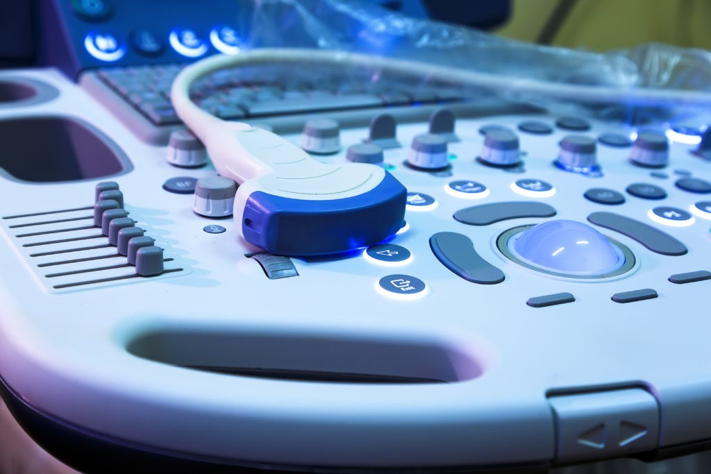
In addition to improved needle quality for imaging, ultrasound technology itself has advanced far enough to significantly facilitate the ease and efficacy of regional anesthesia. Over the past several years, transducer quality, improved image resolution, and better beam formation have all drastically increased the utility of ultrasound in providing accurate sonoanatomy for safe and effective blocks4. In addition, ultrasound technology has ventured into newer modalities that may provide additional ease to performing regional anesthesia; three-dimensional ultrasound imaging can help elucidate anatomic structures easier, facilitate more precise needle movements, and map the spread of local anesthetic around the target area.
The use of ultrasound guidance of regional anesthesia has come a long way, and many more helpful advances are coming into widespread use. Machine learning may play a key role in imaging processing, with nearly complete minimization of artifact and shadowing in order to provide better visualization of structures. In addition, machine learning may eventually become advanced enough to automatically identify specific anatomic structures in real-time to aid the provider in reaching the intended target. Through improvement of beam formation and image resolution to better penetrate structures with poor acoustic transmission, even neuraxial anesthesia can receive immense benefit from ultrasound guidance. The use of automation may also be a key development in the advancement of regional anesthesia, helping providers maneuver the ultrasound probe or needle in order to administer the most precise block possible. The practice of ultrasound-guided regional anesthesia has come so far since its first reported use in 19941; it is extremely likely that in another 25 years, the field will see immense improvements in patient safety, success rates, and utility for numerous clinical situations.
1. Gray AT. Ultrasound-guided regional anesthesia: current state of the art. Anesthesiology. 2006;104(2):368-373, discussion 365A.
2. Kapral S, Greher M, Huber G, et al. Ultrasonographic guidance improves the success rate of interscalene brachial plexus blockade. Reg Anesth Pain Med. 2008;33(3):253-258.
3. Hebard S, Hocking G. Echogenic technology can improve needle visibility during ultrasound-guided regional anesthesia. Reg Anesth Pain Med. 2011;36(2):185-189.
4. Henderson M, Dolan J. Challenges, solutions, and advances in ultrasound-guided regional anaesthesia. BJA Education. 2016;16(11):374-380.
5. Kåsine T, Romundstad L, Rosseland LA, et al. Needle tip tracking for ultrasound-guided peripheral nerve block procedures-An observer blinded, randomised, controlled, crossover study on a phantom model. Acta Anaesthesiol Scand. 2019.
6. Clendenen NJ, Robards CB, Clendenen SR. A standardized method for 4D ultrasound-guided peripheral nerve blockade and catheter placement. Biomed Res Int. 2014;2014:920538.