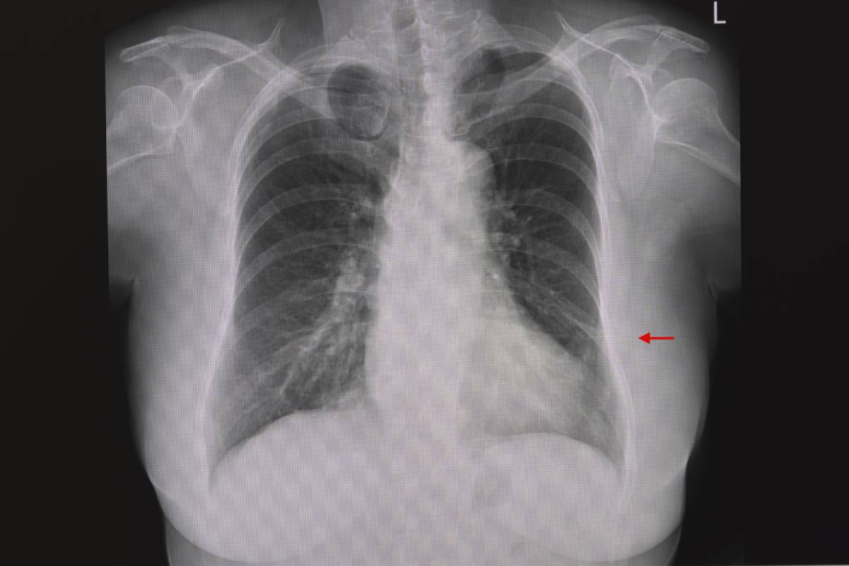
Atelectasis occurs when air is not able to fully expand the alveoli of the lungs. 90% of patients undergoing general anesthesia experience atelectasis (Randtke et al, 2015). This is because general anesthesia decreases muscle tone and thus functional residual capacity (FRC) — the amount of air that is left in the lungs after exhaling. Atelectasis increases the risk for hypoxemia and pneumonia and can continue into the postoperative period. Preventing and reversing atelectasis can improve patient outcomes for surgeries that require general anesthesia (Randtke et al, 2015).
There are three explanations of the physiological cause of atelectasis that are generally agreed upon. The absorption mechanism is based on oxygen balance. Often, patients undergoing anesthesia will be placed on 100% oxygen, which pushes nitrogen gas out of the lungs (nitrogen is a component of normal atmospheric air) (Randtke et al, 2015). Oxygen is absorbed by the capillary bed, leaving the alveoli with too little gas remaining inside. Since the alveoli are not supported by cartilage, only the pressure of gases against their walls, they then collapse (Randtke et al, 2015). This can also occur with a low ventilation to perfusion ratio — when the capillary beds absorb oxygen faster than the person’s breathing can provide more air. The decreased FRC with general anesthesia exacerbates this problem. It is a challenge to get the appropriate oxygen balance, as not providing enough oxygen can also lead to hypoxemia during the procedure (Randtke et al, 2015).
The compression mechanism for atelectasis occurs when the pleural pressure in the chest cavity is greater than the intrapulmonary pressure. With the reduced muscle tone that occurs under anesthesia, the weight of the chest and the patient’s organs against the diaphragm are significant contributing factors in this mechanism (Randtke et al, 2015). Inflammation and buildup of fluid in the pleural space can also contribute. The pressure can push residual air out of the lungs, further decreasing the FRC and leading to alveolar collapse (Randtke et al, 2015).
The third mechanism is related to abnormalities in surfactant. Surfactant is produced by specific cells in the alveoli and reduces surface tension, making it easier for the lungs to expand (Randtke et al, 2015). Reduced surfactant makes it more difficult for alveoli to stay open in the first place, re-inflate once collapsed and stabilized once reopened. General anesthesia may have some impact on surfactant abnormalities but more research is needed to clarify its role in atelectasis in this context (Randtke et al, 2015).
As shown in a recent meta-analysis regarding prevention and treatment pathways of postoperative pulmonary complications, there is not a lot of high-quality data currently available (Odor et al, 2020). With the lower quality data that is available, interventions found to have a significant effect include postoperative continuous positive airway pressure (CPAP), mucolytic medications (specifically ambroxol) and respiratory physiotherapy (a series of muscle training and breathing exercises practiced before and after the surgery). There is moderate quality data for intraoperative lung protective ventilation, defined as using reduced tidal volumes (<8 mL/kg), positive end expiratory pressure of at least 5 cm H2O and intermittent recruitment maneuvers (high pressure applied for a short period of time to inflate the lungs more fully) (Odor et al, 2020). It is also important to look at prevention in specific patient populations, such as in pediatric patients. One newer study suggests that use of CPAP during induction and emergence in children decreases the risk of intraoperative atelectasis, as well as the risk of residual atelectasis in the postoperative period (Acosta et al, 2021). Another recent study suggests that high-flow nasal cannula oxygen in the postoperative period reduces residual atelectasis (Lee et al, 2021). Overall, more high-quality comparative research on the effectiveness of different prevention methods is needed for all patient populations.
References
Acosta CM, Lopez Vargas MP, Oropel F, et al. Prevention of atelectasis by continuous positive airway pressure in anaesthetised children: A randomised controlled study. Eur J Anaesthesiol. 2021;38(1):41-48. doi:10.1097/EJA.0000000000001351
Lee J-H, Ji S-H, Jang Y-E, Kim E-H, Kim J-T, Kim H-S. Application of a High-Flow Nasal Cannula for Prevention of Postextubation Atelectasis in Children Undergoing Surgery: A Randomized Controlled Trial. Anesthesia & Analgesia. 2020;133(2):474-482. doi:10.1213/ane.0000000000005285
Odor PM, Bampoe S, Gilhooly D, Creagh-Brown B, Moonesinghe SR. Perioperative interventions for prevention of postoperative pulmonary complications: systematic review and meta-analysis. BMJ. 2020;368:m540. doi:10.1136/bmj.m540
Randtke MA, Andrews BP, Mach WJ. Pathophysiology and Prevention of Intraoperative Atelectasis: A Review of the Literature. J Perianesth Nurs. 2015;30(6):516-527. doi:10.1016/j.jopan.2014.03.012
