An accurate estimation of the timing of labor is important for several clinical decisions, including determination of preterm birth and necessary treatment regimens [1]. Currently, gestational age and due date is approximated based on information about the last menstrual cycle and assumes a gestational length of 40 weeks [1,2]. Ultrasound imaging in early pregnancy can also be used to determine gestational age and estimated date of delivery [1]. Although these approaches are useful in managing pregnancy, neither method is an accurate predictor of when labor will begin, since most pregnancies deviate from the standard of 40 weeks of gestational duration [2]. To advance clinical decision-making, efforts have been made to better predict the actual onset of labor using predictive biomarkers measured by blood tests [2].

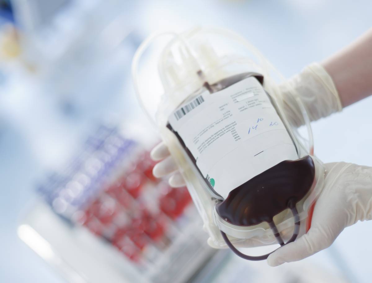
During pregnancy, the maternal circulatory system connects with the fetal circulatory system through the placenta, carrying a variety of compounds such a steroid hormones, micronutrients, and nucleic acids [1]. The maintenance of pregnancy relies on finely tuned adaptions to the concentrations of these biomarkers, which can be easily detected in maternal blood using high-content metabolomic, proteomic, and single-cell cytometric technologies [2].

A 2019 study funded by the University Hospitals of Leicester yielded results that support the idea that it may be possible to predict the onset of labor from a single blood test [3]. The researchers monitored the plasma concentrations of N-arachidonylethanolamine (AEA), N-acylethanolamines (NAE), N-oleoylethanolamide (OEA), and N-palmitoylethanolamide (PEA) in 217 pregnant patients [3]. In line with previous studies that had shown that plasma AEA concentrations increase in the third trimester and peak in labor, they found that women at risk for pre-term labor also had an elevated plasma AEA concentration [3]. The data suggested that a single AEA measurement taken from a blood sample can predict the gestational age of delivery and the remaining duration of pregnancy with better accuracy compared to conventional methods [3].

Similarly, a 2021 study by Stanford University sought to determine the dynamic changes in the maternal metabolome, proteome, and immunome preceding the day of labor [2]. The analysis of the results revealed a marked transition from pregnancy maintenance to pre-labor physiology starting 2 to 4 weeks before labor onset [2]. Endocrine and inflammatory changes were pronounced during late pregnancy, with steroid hormone metabolites being among the most informative biomarkers [2]. A decline in progesterone levels was linked to the progression to labor [2]. Moreover, the surge in steroid hormone metabolites weeks before labor coincided with changes in plasma protein concentrations and immune cell responses [2]. A sharp increase in the concentration of IL-4, an IL-33 antagonist, was found to play a prominent regulatory role during the pre-labor phase [2]. In mice, IL-33 has been shown to have a role in pregnancy maintenance [2].

The non-invasive nature of maternal blood tests offers great promise as a way to predict labor onset [3]. Research continues to determine the best biomarkers for estimating the timing of labor and has the potential to transform the management of pregnancy, especially in relation to pre-term birth [1-3].

References

  1. Liang, L., Rasmussen, M., Piening, B. etc. (2020). Metabolic dynamics and prediction of gestational age and time to delivery in pregnant women. Cell181(7), 1680-1692. doi:10.1016/j.cell.2020.05.002
  2. Stelzer, I., Ghaemi, M., Han, X. etc. (2021). Integrated trajectories of the maternal metabolome, proteome, and immunome predict labor onset. Science Translational Medicine13(592). doi:10.1126/scitranslmed.abd9898
  3. Bachkangi, P., Taylor, A., Bari, M. etc. (2019). Prediction of preterm labour from a single blood test: The role of the endocannabinoid system in predicting preterm birth in high-risk women. European Journal of Obstetrics & Gynecology and Reproductive Biology243, 1-6. doi:10.1016/j.ejogrb.2019.09.029

Distal radius fractures, or breaks in the radius bone of the arm close to the wrist, are among the most common types of bone fractures. Many of these fractures, particularly if they are displaced (i.e., if the bone fragments on either side of the break are not aligned), require surgery, known as distal radius repair, to ensure proper setting and healing of the radius.1 There are several options for anesthesia during radius repair—most notably general anesthesia, local anesthesia, and peripheral nerve blocking (a specific type of local anesthesia)—and recent studies have begun to evaluate the benefits and drawbacks of each approach, particularly with regard to pain control, postoperative functional outcomes, and length of stay.

Some studies have identified better clinical outcomes for distal radius repair with local, rather than general, anesthesia. For instance, Egol et al. collected data from 187 patients and found that patients who received local anesthesia had improved wrist and finger range of motion compared with patients who received general anesthesia and also showed higher functional scores (as measured by Disabilities of the Arm, Shoulder and Hand) at 3- and 6-month follow-ups.2 In contrast, Rundgren et al. found using a single-center randomized clinical trial of 88 patients that, while local anesthesia appeared to significantly reduce early patterns of postoperative pain and opioid consumption after radius repair, neither total opioid consumption nor longer-term functional outcomes differed significantly between the local and general anesthesia groups.3

Peripheral nerve blocking, a type of local anesthesia, has also recently been employed in comparison studies with general anesthesia to examine its effects on outcomes following distal radius repair. For example, Galos et al. conducted a randomized controlled study of 36 patients to answer four questions: (1) whether patients receiving general anesthesia or brachial plexus blockade (a type of peripheral nerve blockade administered through a single infraclavicular shot) had worse pain scores at various time points, (2) whether there was a difference in operating suite time and recovery room time between the two groups, (3) whether either group experienced higher postoperative narcotic use, and (4) whether either group displayed higher functional assessment scores 6 and 12 weeks after surgery. The researchers found that patients who received general anesthesia had worse pain 2 hours postoperatively, while patients who received a brachial plexus blockade reported worse pain 12 hours postoperatively, suggesting that “rebound pain” ought to be taken into consideration when employing peripheral nerve blocking. Both time in the recovery room and overall amount of narcotics consumed was higher for patients who received general anesthesia, but functional scores did not differ between the two groups.4

In addition, Johnson et al. found this past year, based on a review of 80 patients, that patients who were treated with peripheral nerve blocking reported a significant decrease in postoperative pain at discharge as well as decreased length of stay. These researchers reported one minor complication with peripheral nerve blocking: a short-lived skin irritation at the site of injection.5

References 

(1)  Distal Radius Fracture (Wrist Fracture) | Johns Hopkins Medicine https://www.hopkinsmedicine.org/health/conditions-and-diseases/distal-radius-fracture-wrist-fracture

(2)  Egol, K. A.; Soojian, M. G.; Walsh, M.; Katz, J.; Rosenberg, A. D.; Paksima, N. Regional Anesthesia Improves Outcome After Distal Radius Fracture Fixation Over General Anesthesia. Journal of Orthopaedic Trauma 201226 (9), 545–549. https://doi.org/10.1097/BOT.0b013e318238becb. 

(3)  Rundgren, J.; Mellstrand Navarro, C.; Ponzer, S.; Regberg, A.; Serenius, S.; Enocson, A. Regional or General Anesthesia in the Surgical Treatment of Distal Radial Fractures: A Randomized Clinical Trial. The Journal of Bone and Joint Surgery 2019101 (13), 1168–1176. https://doi.org/10.2106/JBJS.18.00984. 

(4)  Galos, D. K.; Taormina, D. P.; Crespo, A.; Ding, D. Y.; Sapienza, A.; Jain, S.; Tejwani, N. C. Does Brachial Plexus Blockade Result in Improved Pain Scores After Distal Radius Fracture Fixation? A Randomized Trial. Clinical Orthopaedics & Related Research 2016474 (5), 1247–1254. https://doi.org/10.1007/s11999-016-4735-1. 

(5)  Johnson, P.; Hustedt, J.; Matiski, T.; Childers, R.; Lederman, E. Improvement in Postoperative Pain Control and Length of Stay With Peripheral Nerve Block Prior to Distal Radius Repair. Orthopedics 202043 (6). https://doi.org/10.3928/01477447-20200721-14. 

COVID-19, like many viral diseases, continues to impact patients’ health even after they have resolved their initial infections. Symptoms manifest across different organ systems with varying levels of severity. A literature review published March 22, 2021 in Nature Medicine by Nalbandian et al. surveyed the current research on post-acute COVID-19 disease. Across different studies of post-acute sequelae of SARS-CoV-2 infection (PASC), researchers observed pulmonary, cardiovascular, neuropsychiatric, hematologic, renal, endocrine, gastrointestinal, and dermatologic conditions, underscoring the complex, heterogeneous nature of the syndrome.1

In a prospective cohort study of 1,733 Chinese patients observed at six months from the onset of COVID-19 symptoms, Huang et al. found that a majority of the patients (76 percent) reported at least one PASC symptom. Fatigue or muscular weakness were the most commonly reported symptom (63 percent), in line with other studies.2 Chronic fatigue has been observed after numerous other acute infections, like SARS coronavirus.3 In one analysis, nearly all (97 percent) of a cohort of 29 patients admitted to a rehabilitation center after overcoming severe COVID-19 still experienced gait speed deficits at discharge from the rehabilitation center.4

Pulmonary complications are notably prevalent as well, with over one-fifth (23 percent) of participants in the study by Huang et al. experiencing dyspnea.2 In smaller studies conducted over shorter follow-up periods, this number was as high as 40 percent. Other observed symptoms include cough and persistent oxygen requirements. Those who had more severe acute COVID-19, including patients who required a high-flow nasal cannula or mechanical ventilation, may be at risk for serious long-term complications like pulmonary fibrosis and pneumonia.1

Neuropsychiatric complications continue to impact the quality of life and daily activities of a significant number of COVID-19 survivors. In the study by Huang et al., 23 percent of individuals experienced anxiety or depression and 26 percent had sleep difficulties.2 In a small United Kingdom study, 30 percent of hospitalized COVID-19 patients had symptoms of PTSD. In all of the studies summarized by Nalbandian et al., loss of taste and smell was experienced by over 10 percent of study participants. Cognitive impairment has widely been noted as well, with many suffering from “brain fog” (difficulties with concentration, memory, executive function and more).1

Beyond impacting individuals’ wellbeing, cognitive and physical fatigue associated with post-acute COVID-19 will alter productivity and the economy. Chronic fatigue symptoms bear resemblance to myalgic encephalomyelitis/chronic fatigue syndrome (ME/CFS), which currently poses an estimated financial burden of between $17 and $24 billion in the U.S. each year. Experts propose that even if only 25 million Americans contract COVID-19 by the end of 2021 and only 10 percent subsequently suffer from an illness meeting the ME/CFS definition, the number of Americans suffering from ME/CFS would at least double over one year. Globally, they estimate that the number of people suffering from ME/CFS would increase to 110 million during 2021.3

Pulmonary complications will also pose economic consequences. While only one year of data is available thus far on the healthcare costs associated with COVID-19, the Kaiser Family Foundation studied these figures to estimate that the annual cost of treating COVID-19 cases only for uninsured Americans might range from $13.9 billion to $41.8 billion.5 It is also necessary to consider healthcare costs for managing conditions that can be exacerbated by lasting damage from a previous COVID-19 infection, such as pneumonia. Before the pandemic, roughly 1.5 million people were hospitalized for pneumonia each year in the U.S., at an average cost of $20,000 per stay.

The novelty of COVID-19 and the limited opportunities to observe its ongoing impacts mean that much is unknown about the prevalence and characteristics of post-acute conditions. Hypotheses about their causes abound: the effects of hospitalization, acute respiratory distress syndrome, and the impact of the hyperinflammatory response associated with COVID-19 may all play roles. Other important factors are confounding health disparities, social determinants of health, and the psychosocial impact of the pandemic.6

In December, Congress promised $1.15 billion over four years for the National Institutes of Health to fund research on the prolonged health consequences of COVID-19.7 Moreover, over 30 U.S. hospitals and health systems have already established clinics solely devoted to post-COVID-19 research and care.8 The long-term effects of COVID-19 remain unclear, and health systems, researchers, and clinicians are still in the initial stages of learning about how to best care for patients.

References 

  1. Nalbandian A, Sehgal K, Gupta A, et al. Post-acute COVID-19 syndrome. Nat Med. 2021;27(4):601-615. 
  1. Huang C, Huang L, Wang Y, et al. 6-month consequences of COVID-19 in patients discharged from hospital: a cohort study. Lancet. 2021;397(10270):220-232. 
  1. Komaroff AL, Bateman L. Will COVID-19 lead to myalgic encephalomyelitis/chronic fatigue syndrome? Front Med (Lausanne). 2020;7:606824. 
  1. Olezene CS, Hansen E, Steere HK, et al. Functional outcomes in the inpatient rehabilitation setting following severe COVID-19 infection. PLoS One. 2021;16(3):e0248824. 
  1. Dovere E-I. Vaccine refusal will come at a cost—for all of us. Atl Mon. Published online April 10, 2021. https://www.theatlantic.com/politics/archive/2021/04/vaccine-refusal-hesitancy-economic-costs/618528/ 
  1. Bhadelia, N. “Post Acute Sequelae of SARS-CoV-2 (PAS-C)” Lecture presented at MassCPR Scientific Symposium: COVID-19 Diagnostic Testing and Clinical Management; March 30, 2021; https://www.youtube.com/watch?v=5RX2IM4Srgc.  
  1. NIH launches new initiative to study “Long COVID.” NIH. Published February 23, 2021. https://www.nih.gov/about-nih/who-we-are/nih-director/statements/nih-launches-new-initiative-study-long-covid 
  1. Carbajal E. 30 hospitals, health systems that have launched post-COVID-19 clinics. Beckers Hospital Review. https://www.beckershospitalreview.com/patient-safety-outcomes/13-hospitals-health-systems-that-have-launched-post-covid-19-clinics.html

The field of outpatient services has grown rapidly over the last several years, spurred by changes to reimbursements for inpatient procedures from health plan providers. One result of this is an increasing number of hybrid surgery centers — facilities that operate as both an office-based laboratory (OBL) and an ambulatory surgical center (ASC). Usually, a hybrid suite contains the imaging equipment typical of an OBL, as well as the equipment and sterilization standards necessary for surgical and non-surgical interventions [1]. While hybrid surgery centers can offer advantages to both patients and healthcare providers that a traditional hospital setting cannot, the transition to this structure may prove difficult. OBL-ASC facilities are complex structures that have many financial, legal, and practical hurdles.  

The flexibility of hybrid suites is advantageous to medical organizations and patients alike. The ability to transition from office to OR can improve patient outcomes in the event of an emergency during a routine procedure [2]. It also allows for non-surgical and surgical procedures to be performed consecutively or simultaneously, thus reducing the number of hospital admissions and shortening the length of stay for patients, which saves money and increases efficiency [3]. Hybrid centers have the potential to guide the standard for medical treatment toward a more integrated approach [4]. Improving patient outcomes and satisfaction is also beneficial for medical providers as the industry shifts toward value-based care [5]. Structurally, the hybrid center is less bureaucratic than the traditional hospital and allows physicians to increase their income through equity ownership [6].  

Despite these prospective benefits, there are many logistical obstacles to instituting a hybrid surgery center. Legal regulations can make it difficult to build a hybrid suite or convert existing offices. Under federal law, in order for an OBL and ASC to operate within the same facility, they must maintain different hours of operation and keep medical records separate. Certain states also restrict the proliferation of outpatient clinics based on need and access [7]. The performance of hybrid procedures must be accommodated not only by new facilities but by new, interdisciplinary working dynamics. Optimizing the design of hybrid centers will require repeated study of the workflow in these environments, and improved collaboration between medical professionals [7].  

Despite the difficulties of establishing hybrid surgery centers, these facilities are growing rapidly and encompass a large share of healthcare services in the country. This trend is the result of a confluence of factors. Many insurance providers no longer cover routine procedures when performed in a hospital because of the higher costs associated with inpatient procedures [8]. Patients, too, object to high costs and have demanded alternatives, pressuring the industry to embrace hybrid centers [5]. The most recent development accelerating the shift, however, is more urgent than market forces. In order to minimize exposure and free up hospital resources for COVID-19 patients, pressure for outpatient centers to increase their caseload has grown. While the pandemic is ongoing, outpatient facilities, including hybrid centers, are easing the strain on hospitals by providing care that would otherwise have been delayed [9].       

References 

[1] Bazzi, May, et al. “The Drama in the Hybrid OR: Video Observations of Work Processes and Staff Collaboration During Endovascular Aortic Repair.” Journal of Multidisciplinary Healthcare, vol. 12, 2019, pp. 453-464, doi: 10.2147/JMDH.S197727.   

[2] “Hybrid Surgical Procedures.” Hybrid Operating Rooms & Hybrid Cath Labs, J.M. Keckler Medical Co., hybridoperatingroom.com/hybrid-surgical-procedures/. 

[3] Bazzi, May, et al. “Team Composition and Staff Roles in a Hybrid Operating Room: A Prospective Study Using Video Observations.” Nursing Open, 2019, doi: 10.1002/nop2.327.   

[4] Davidson, Michael J. and Tsuyoshi Kaneko. “Use of the Hybrid Operating Room in Cardiovascular Medicine.” Circulation, vol. 130, no. 11, 2014, pp. 910-917, doi: 10.1161/CIRCULATIONAHA.114.006510

[5] Derek Long, David McMillan, et al.| CPA, ASA | Apr 1, 2019. “HOPDs vs. ASC: Understanding Payment Differences.” 1 Apr. 2019, https://www.hfma.org/topics/hfm/2019/ april/hopds-vs–asc–understanding-payment-differences.html. 

[6] Tim van Biesen and Todd Johnson | Healthcare Private Equity Advisers | Sept 23, 2019. “Ambulatory Surgery Center Growth Accelerates: Is Medtech Ready?” Bain & Company, 23 Sept. 2019, https://www.bain.com/insights/ambulatory-surgery-center-growth-accelerates-is- medtech-ready/. 

[7] Cilek, Jacob A. and Jason S. Greis. 12 Business and Legal Considerations for Developing a ‘Hybrid’ Office-Based Laboratory–Ambulatory Surgery Center. McGuire Woods, Sept. 2019, https://media.mcguirewoods.com/publications/2019/OBL-ASC-Hybrid-Article-12-Considerations.pdf. 

[8] Karen Blum | Office of Johns Hopkins Physicians | Apr 25, 2018. “Shifting Low-Risk Procedures to Ambulatory Surgery Centers.” BestPractice News, 25 Apr. 2018, https://www. hopkinsmedicine.org/office-of-johns-hopkins-physicians/best-practice-news/shifting-low-risk-procedures-to-ambulatory-surgery-centers. 

[9] Jacqueline LaPointe | Revcycle Intelligence | Mar 17, 2020. “Hospitals Delay, Shift Surgeries to Outpatient Due to COVID-19.” Revcycle Intelligence, 17 Mar. 2020, https://revcycleintelligen ce.com/news/hospitals-delay-shift-surgeries-to-outpatient-due-to-covid-19.

Anemia is a condition that results in low red blood cell count and hemoglobin deficiency, decreasing the blood’s capacity to carry oxygen [1]. The World Health Organization specifies that hemoglobin concentrations of less than 12.0g/dl in non-pregnant women and 13.0g/dl in men could be considered anemic [2]. There are a number of potentially serious consequences of anemia, including increased cardiac output, which can lead to damage to the heart’s muscular tissue [3]. Perioperative anemia is not uncommon and can make surgery and recovery significantly more complicated. Furthermore, anemia in surgical patients is associated with higher morbidity and mortality rates [4].  

In order to improve the survival and recovery outcomes for anemic surgical patients, the condition must first be diagnosed and treated appropriately. Unfortunately, perioperative anemia often does not receive the attention it warrants until hemoglobin levels become low enough for blood transfusion [5]. A study by Beattie et al. found that transfusion rates were three times higher for anemic patients than their non-anemic counterparts. [2]. When transfusions are required, the risk of complications such as ischemic stroke and myocardial infarction rises significantly, and transfusions are associated with higher morbidity rates among surgical patients [6].  

As anemia has gained attention in the medical community, a number of recommendations and strategies for preventing morbidities associated with anemia in surgical patients have been released. In 2005, an interdisciplinary panel of medical professionals advised that elective surgery patients have their hemoglobin levels tested at least thirty days before surgery and that unexplained anemia should always be considered as an effect of a condition that requires pre-surgical attention [7]. The American Society of Anesthesiology advises that the necessity for blood transfusion is usually indicated at a hemoglobin level of <6g/dl, but that ischemia, bleeding, and other risks should be weighed alongside it [6]. More recently, researchers created an automated system for iron-deficiency anemia screening, which they found to improve diagnosis over clinical procedures [4].  

Concerns regarding morbidity rates for anemic patients extend to all members of the surgical team, including anesthesia providers. The anesthesia provider should review vital signs and patient data and evaluate whether red blood cell transfusion is worth the risk to the patient. Because anemia is associated with poorer outcomes, care providers must carefully weigh the decision to administer a transfusion [1]. Several medical associations have issued guidelines for evaluating the necessity of transfusion. These guidelines include considerations of hemoglobin levels as well as various health factors. Current trends indicate that physicians’ ability to identify and treat anemia is developing rapidly, improving outcomes for surgical patients with anemia. 

References 

[1] Klick, John C., and Edwin G. Avery. “Anesthetic Considerations for the Patient With Anemia and Coagulation Disorders.” Anesthesiology, edited by David E. Longnecker et al, McGraw-Hill, 2012, pp. 196-216.   

[2] Beattie, Scott W., et al. “Risk Associated With Preoperative Anemia in Noncardiac Surgery: A Single-Center Cohort Study.” Anesthesiology, vol. 110, 2009, doi: 10.1097/ALN.0b013e318 19878d3. 

[3] “Anemia Compensation.” Open Anesthesia, 26 Feb., https://www.openanesthesia.org/anemia _compensation/

[4] Okocha, Obianuju, et al. “An Effective and Efficient Testing Protocol for Diagnosing Iron-Deficiency Anemia Preoperatively.” Anesthesiology, vol. 133, 2020, 109-118, doi: 10.1097/ALN.0000000000003263. 

[5] Warner, Matthew A., et al. “Perioperative Anemia: Prevention, Diagnosis, and Management Throughout the Spectrum of Perioperative Care.” Anesthesia & Analgesia, vol. 130, no. 5, 2020, 1364-1380, doi: 10.1213/ANE.0000000000004727. 

[6] Shander, Aryeh et al., “Anesthesia for Patients With Anemia.” Anesthesiology Clinics, vol. 34, no. 4, 2016, 711-730, doi: 10.1016/j.anclin.2016.06.007. 

[7] Goodnough, Laurence T., et al. “Detection, Evaluation, and Management of Anemia in the Elective Surgical Patient.” Anesthesia & Analgesia, vol. 101, no. 6, 2005, 1858-1861, doi: 10.1213/01.ANE.0000184124.29397.EB. 

The SARS-CoV-2 VUI 202012/01 variant sits poised to become the dominant coronavirus variant in the U.S. as early as March.1 Scientists first sequenced the variant, also known as B.1.1.7, in the U.K. in September 2020.2 It quickly garnered scientific and public attention as experts hypothesized that B.1.1.7’s fast spread contributed to the case spike in England. Recent research – yet to undergo peer review – estimates that B.1.1.7’s transmissibility exceeds that of prior variants by 50-75%.3,4 As of January 12, 2021, B.1.1.7 has appeared already in at least 33 countries.2 A key question is how available vaccines affect this SARS-CoV-2 variant.

B.1.1.7 differs from SARS-CoV-2 by 23 mutations in its genetic blueprint.2 Six of the mutations do not cause any change in the amino acid sequence. Eight changes, however, affect the spike protein of the virus. The spike protein plays a key role in receptor recognition and cell membrane fusion; it is this protein that locks onto compounds in the human body to enable infection. Currently, the N501Y mutation is of primary interest to scientists. This mutation – the change of an asparagine to a tyrosine – alters the receptor binding domain of the spike protein.2,5 Scientists hypothesize that this mutation allows the virus to attach more strongly to cells, which, in turn, increases its transmissibility. It is critical to understand whether this SARS-CoV-2 variant is different enough as to affect the efficacy of available vaccines.

Current vaccines target these spike proteins to impede the virus’s ability to latch onto host cells.2 A person’s immune response to the vaccine produces antibodies that bind to various locations on the spike protein in order to neutralize the virus. Experts fear, however, that certain mutations to the spike protein may destroy or impair the sites where antibodies bind.6 Fortunately, the N501Y mutation’s location makes it such that the mutation likely does not significantly affect antibody binding sites.6,7  

Currently, the CDC reports that “there is no evidence to suggest that the variant [B.1.1.7] has any impact on the severity of disease or vaccine efficacy.”8 In a January 7 preprint, scientists at Pfizer and the University of Texas Medical Branch at Galveston concluded that the N501Y mutation did not affect the Pfizer vaccine-generated antibodies’ ability to latch on to the virus.9 In this study, researchers examined whether the sera of Phase 3 trial participants neutralized the B.1.1.7 variant as well as they neutralized the non-B.1.1.7 virus.10 The results suggest the N501Y mutation does not confer Pfizer vaccine resistance because the same quantity of serum successfully neutralized both the original and the mutated virus. Likewise, the Moderna vaccine appears to confer immunity that recognizes B.1.1.7. Early laboratory tests looked at blood samples from eight people and two primates who received the two Moderna vaccine doses. The scientists observed no decrease in neutralization capacity in the serum against B.1.1.7.11 In addition, Novavax reports its vaccine efficacy as 85.6% protective against B.1.1.7 in Phase 3 UK clinical trials.12  

Neither Oxford-AstraZeneca nor Johnson & Johnson have yet released data that specifically examines their vaccines’ efficacy against B.1.1.7. Oxford-AstraZeneca scientists currently have an analysis underway that examines the vaccine’s neutralization capacity against the variant; they expect to report the results within two weeks.13 Moreover, recently released data from Johnson & Johnson claim 66% vaccine efficacy against COVID-19 infection and 85% efficacy against severe cases; unfortunately, the data does not speak explicitly to the vaccine’s efficacy against B.1.1.7.14 However, the study spanned multiple continents and concluded relatively recently, which makes it likely that some participants were exposed to B.1.1.7.  

At present, scientists seem optimistic that current vaccines will confer some level of protection against the B.1.1.7 SARS-CoV-2 variant. As new variants arise, however, the current vaccines will continue to be tested. 

References 

1. Branswell, H. Coronavirus variant could become dominant strain by March, CDC warns. STAT News https://www.statnews.com/2021/01/15/covid19-b117-variant-cdc/ (2021). 

2. Cohut, M. & Hewnigs-Martin, Y. New coronavirus variant: What we know so far. https://www.medicalnewstoday.com/articles/covid-19-what-do-we-know-about-the-new-coronavirus-variant (2021). 

3. Davies, N. G. et al. Estimated transmissibility and severity of novel SARS-CoV-2 Variant of Concern 202012/01 in England. medRxiv (2020) doi:10.1101/2020.12.24.20248822. 

4. Volz, E. et al. Transmission of SARS-CoV-2 Lineage B.1.1.7 in England: Insights from linking epidemiological and genetic data. medRxiv (2021) doi:10.1101/2020.12.30.20249034. 

5. Rathnasinghe, R. et al. The N501Y mutation in SARS-CoV-2 spike leads to morbidity in obese and aged mice and is neutralized by convalescent and post-vaccination human sera. medRxiv (2021) doi:10.1101/2021.01.19.21249592. 

6. Moore, J. P. & Offit, P. A. SARS-CoV-2 Vaccines and the Growing Threat of Viral Variants. JAMA (2021) doi:10.1001/jama.2021.1114. 

7. Starr, T. N. et al. Deep Mutational Scanning of SARS-CoV-2 Receptor Binding Domain Reveals Constraints on Folding and ACE2 Binding. Cell 182, 1295-1310.e20 (2020) doi: 10.1016/j.cell.2020.08.012. 

8. CDC. Emerging SARS-CoV-2 Variants. Centers for Disease Control and Prevention https://www.cdc.gov/coronavirus/2019-ncov/more/science-and-research/scientific-brief-emerging-variants.html (2020). 

9. Xie, X. et al. Neutralization of N501Y mutant SARS-CoV-2 by BNT162b2 vaccine-elicited sera. bioRxiv (2021) doi:10.1101/2021.01.07.425740. 

10. Pfizer. An In Vitro Study Shows Pfizer-BioNTech COVID-19 Vaccine Elicits Antibodies that Neutralize SARS-COV-2 with a Mutation Associated with Rapid Transmission. News | Pfizer https://www.pfizer.com/news/press-release/press-release-detail/vitro-study-shows-pfizer-biontech-covid-19-vaccine-elicits (2021). 

11. Wu, K. et al. mRNA-1273 vaccine induces neutralizing antibodies against spike mutants from global SARS-CoV-2 variants. bioRxiv (2021) doi:10.1101/2021.01.25.427948. 

12. Novavax, Inc. Novavax COVID-19 Vaccine Demonstrates 89.3% Efficacy in UK Phase 3 Trial. 3 https://ir.novavax.com/node/15506/pdf (2021). 

13. Rivas, K. AstraZeneca expects COVID-19 vaccine data on UK variant within 2 weeks. Fox News https://www.foxnews.com/health/astrazeneca-expects-covid-19-vaccine-data-uk-variant-within-2-weeks (2021). 

14. Johnson & Johnson. Johnson & Johnson Announces Single-Shot Janssen COVID-19 Vaccine Candidate Met Primary Endpoints in Interim Analysis of its Phase 3 ENSEMBLE Trial. https://www.jnj.com/johnson-johnson-announces-single-shot-janssen-covid-19-vaccine-candidate-met-primary-endpoints-in-interim-analysis-of-its-phase-3-ensemble-trial (2021).    

Post-operative sore throat (POST) occurs in over half of the patients who undergo general anesthesia with tracheal intubation every year1. While POST is considered a mild post-operative complication with little long-term effect on patient outcome, it does contribute to post-operative morbidities and decrease patient satisfaction1.  

Much of the literature on the causes of post-operative sore throat center around a set of key risk factors. Female sex and young age are commonly named, as well as not administering a neuromuscular block, doing nasal intubation instead of oral, high endotracheal cuff pressures, and laryngeal mucosal injury2. A recent study found a correlation between the size of endotracheal tube used and the incidence of POST complaints from patients, stating that a tube of size 6.5mm or bigger significantly increased risk of POST2. The article reasoned that a bigger tube may be causing direct trauma to the tracheal mucosa, resulting in the release of inflammatory cytokines that produce those tell-tale sore throat symptoms. Clinicians can take measures to reduce the risk of POST, using the smallest possible tube, monitoring cuff pressures, and being extra careful with women and children who are at higher risk2.   

Since two of the major risk factors involve the use of endotracheal tubes, there has been an increased interest in finding ways to avoid using endotracheal tubes. One method with considerable success is using a supraglottic airway device, such as the laryngeal mask airway3. A 2020 study in BMC Anesthesiology showed that using a flexible, reinforced laryngeal mask airway reduced the incidence and the severity of POST after thyroid surgery when compared to an endotracheal tube, as well as better hemodynamics during intubation and less buckling during extubation3. These findings are likely due to the positioning of the LMA, which is placed superior to the larynx, causing less mucosal trauma to the trachea, and the flexible nature of the LMA, which decreases cuff pressures3.  

Furthermore, there has been some investigation in the past five years into pharmacological prevention techniques, such as steroids, NSAIDs, NMDA receptor agonists and even licorice1. Steroid interventions such as IV dexamethasone, the application of triamcinolone paste on the tracheal tube, betamethasone gel and pre-operative inhaled fluticasone reduced the likelihood and severity of POST, so long as tube size and cuff pressures are also controlled1. Neither NSAIDs nor licorice were effective 24 hours after surgery, but they did alleviate some POST symptoms immediately after surgery1. NMDA receptor agonists had variable efficacies, with the magnesium gargle being more effective than the ketamine gargle1.  

These interventions have been effective at reducing the incidence and severity of POST to some degree, but with varying success rates and therapeutic profiles. However, a new prevention technique has recently shown promise: preoperative gum chewing4. In a study published in Anesthesia and Analgesia, a randomized control trial found that chewing gum for two minutes before the administration of general anesthesia and intubation via a supraglottic airway device greatly reduced incidence of both moderate and severe post-operative sore throat4. If chewing gum can be shown to be consistently effective, it could be implemented as a simple way of alleviating patient discomfort with few to no side effects or contraindications4.

References 

  1. El-Boghdadly K, Bailey CR, Wiles MD: Postoperative sore throat: a systematic review. Anaesthesia 2016; 71(6). https://doi.org/10.1111/anae.13438 
  1. Fenta E, Tesfaw A: Incidence and factors associated with postoperative sore throat for patients undergoing surgery under general anesthesia with endotracheal intubation and Debre Taylor General Hospital, North Central Ethiopia: a cross sectional study. International Journal of Surgery: 25: 1-5. https://doi.org/10.1016/j.ijso.2020.06.003 
  1. Gong Y, Xu X, Wang J, Che L, Wang W, Yi J: Laryngeal mask airway reduces incidence of postoperative sore throat after thyroid surgery compared with endotracheal tube: a single-blinded randomized control trial. BMC Anesthesia 2020. https://doi.org/10.1186/s12871-020-0932-2
  1. Wang T, Wang Q, Haiyang Z, Huang S: Effects of preoperative gum chewing on sore throat after general anesthesia with supraglottic airway device: a randomized control trial. Anesthesia and Analgesia 2020; 131 (6): 1864-1871. https://doi.org/10.1213/ANE.0000000000004664 

In 1992, Medicare established a standardized payment scale called the Resource-based Relative Value Scale (RBRVS). While the system succeeded in standardizing rates, it also led to lower payments for some specialties. Anesthesia, in particular, suffered from a rate cut of nearly 30% between 1991 and 1992. Since then, Medicare payments for anesthesia services have amounted to around 33% of commercial rates.[1]

The original RBRVS was based on research by Hsiao et al. which relied heavily on cross-specialty comparisons for similar services. The study looked at three factors: work, practical expense, and professional liability insurance. In general, the researchers compared between 12 and 15 services. However, Hsiao and his team compared just 3 anesthesia services, which accounted for a fraction of the total services offered by anesthesiologists.[2] Ultimately, the researchers decided that anesthesiology services were overvalued by about 41%. As a result, the Medicare rates for anesthesia services were set at a markedly lower rate than other, similar services. 

Between 1995 and 2005, the American Society of Anesthesiologists conducted three studies, each of which showed that Medicare rates for anesthesiology were consistently lower than rates for other specialties. The 1995 and 2000 studies both examined the estimated time required to complete a procedure and found it to be significantly undervalued for anesthesia services. 

The duration of work performed by a medical specialist is one of the primary determinants for payment under the Medicare fee structure. However, studies have shown that physicians consistently overestimate the amount of time required for a procedure.[3] Anesthesia, however, is routinely undervalued. A 2000 study by the ASA estimated that anesthesia times were undervalued by between 28.4% and 37.6% in work Relative Value Units. 

Indeed, some researchers point to the complexity of the RBRVS model as a failing in and of itself. Cooper and Kramer analyzed the RBRVS model and concluded that it was more complex and less effective version compared to other revenue-based cost assignment models.[4] The RBRVS system supposes that all procedures and providers earn the same profit margin, regardless of the scale or complexity of the medical environment. This can translate into standard payments for services that do not correspond to the time put in by the specialist.  

The impact of Medicare rates resonates far beyond the system, as well. The RBRVS model has become more popular with third-party payers as they move from discount-based models to fee schedules. These payers, which include HMOs, use a multiplier linked to the Medicare RBRVS rate to assess their fees. According to Lubarsky and Reves, this results in substantially lower payments for anesthesiologists, not only from Medicare but also from many third-party payers.[5] Indeed, the authors’ study of two different HMOs found that, if Medicare-based multipliers are used, anesthesiologists would need to be paid roughly three times the Medicare rate in order to remain rate-similar with other specialties. 

For almost thirty years, Medicare rates for anesthesia services have remained around 30% lower than similar services offered by other specialties. Multiple studies have demonstrated that this discrepancy is due to shortcomings in the model that establishes the Medicare fee schedule. The effects of these lower payments are felt beyond Medicare—third-party payers that use Medicare rates to set their own fee schedules also routinely underpay anesthesiologists for their services. 

References 

[1] Pregler, Johnathan, et al. “The 33% Problem: Origins and Actions Committee on Economics 33% Workgroup Report ASA Economic Strategic Plan Initiative—October 2020.” ASA Monitor, vol. 84, no. 12, 2020, pp. 28–33., doi:10.1097/01.asm.0000724064.38204.e5

[2] Hsiao, William C. “Resource-Based Relative Values.” JAMA, vol. 260, no. 16, 1988, p. 2347., doi:10.1001/jama.1988.03410160021004.  

[3] Burgette, Lane F., et al. “Estimating Surgical Procedure Times Using Anesthesia Billing Data and Operating Room Records.” Health Services Research, vol. 52, no. 1, 2016, pp. 74–92., doi:10.1111/1475-6773.12474

[4] Cooper, Robin, and Theresa R Kramer. “RBRVS Costing: the Inaccurate Wolf in Expensive Sheep’s Clothing.” Journal of Health Care Finance, vol. 34, no. 3, 2008, pp. 6–18., PMID: 18468375.  

[5] Lubarsky, David A, and J.Gerald Reves. “Using Medicare Multiples Results in Disproportionate Reimbursement for Anesthesiologists Compared to Other Physicians.” Journal of Clinical Anesthesia, vol. 12, no. 3, 2000, pp. 238–241., doi:10.1016/s0952-8180(00)00135-5.

Over the past year, COVID-19’s global spread has continued, leaving no corners of the world untouched. An unprecedented danger, this coronavirus disease has led to more than 1.2 million deaths worldwide and shows no signs of stopping [1]. One major contributing factor to its spread is the multitude of asymptomatic or minimally symptomatic carriers [1]. Despite the severe discomfort that symptomatic patients report experiencing, asymptomatic/minimally symptomatic people lack such symptoms [1]. This absence of pain keeps them from knowing that they carry the disease, increasing the risk that they will infect others [1]. To investigate why people respond to the disease differently and how to reduce infection rates, scientists must study the pathway SARS-CoV-2 follows in the body. Recent research suggests that the neuropilin-1 receptor may be a key piece of the story. 

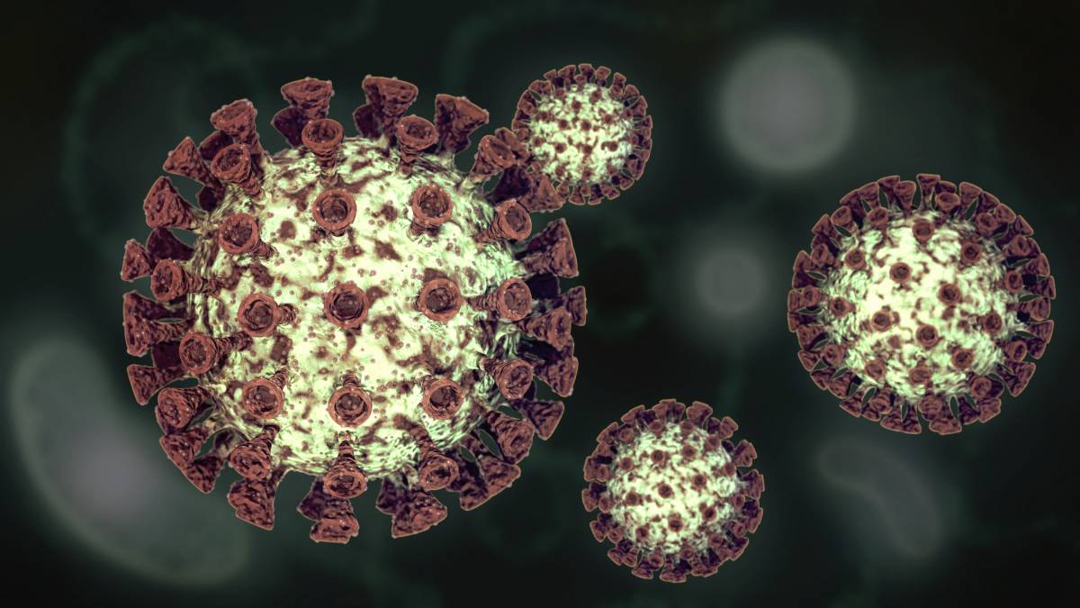
Upon entry into the body, SARS-CoV-2 must enter host cells to prompt viral infection [2]. A spike protein on the outer surface of the virus’ body enables it to attach to human cell receptors [2, 3]. The spike protein can attach itself to the human ACE2 receptor (hACE2) through its receptor-binding domain [2]. Once the virus’ spike protein is bound to hACE2, activation occurs due to human proteases, and the virus can now infect human cells [2]. Up until recently, this was the accepted primary pathway that scientists believed enabled SARS-Cov-2 to infect human cells.  

Recently, scientists have discovered a second pathway through which SARS-CoV-2 infects cells. X-ray crystallography revealed that SARS-CoV-2 can also bind itself to neuropilin-1 (NRP-1) receptor proteins [3]. In cases where the virus attached to both hACE2 and NRP-1, the virus was able to infect many more cells [3]. Conversely, when the spike protein cannot bind to NRP-1, rates of infection significantly decrease–and may, according to one experiment, be nonexistent altogether [3]. The importance of NRP-1 would help explain the virus’s effects on the pulmonary system where NRP1 is abundantly expressed in cells but ACE-2 is almost nonexistent [4]. 

These findings have two prominent implications. First, this knowledge could assist researchers in developing COVID-19 therapies [5]. When spike proteins interact with human proteases, they are cleaved into two polypeptides, S1 and S2 [5]. Scientists believe that this division creates a terminal sequence on S1, which enables the virus to bind to NRP-1 receptors [5]. In SARS-CoV-2 viruses that lack the S1/S2 cleavage site, hamster models have demonstrated a decreased risk of infection [5]. Therefore, if scientists can figure out how to disrupt this cleavage, scientists may discover a meaningful therapy for COVID-19 [5]. 

Additionally, neuropilin-1 may have analgesic effects that explain the lack of pain experienced by asymptomatic and minimally symptomatic COVID-19 patients [1]. The pain felt by symptomatic patients is hypothesized to emanate, at least partially, from SARS-CoV-2-induced cell damage [1]. Researchers discovered that blocking the ligand VEGF-A from binding with NRP-1 inhibited pain perception [1]. They then hypothesized that the interference of the spike protein in NRP-1 signaling inhibits pain during the early stages of COVID-19 [1]. This could be a powerful explanation for why the asymptomatic/minimally symptomatic lack pain, although more research will need to be conducted. 

The power of this secondary infection pathway should not be underestimated. By learning about how NRP-1 signaling is an alternative route to SARS-CoV-2 infection, researchers may find a way to control this novel disease and also better understand the physiological mechanisms of pain. 

References 

[1] A. Moutal et al., “SARS-CoV-2 Spike protein co-opts VEGF-A/Neuropilin-1 receptor signaling to induce analgesia,” Pain, Oct 2020. [Online]. Available: https://doi.org/10.1097/j.pain.0000000000002097 

[2] J. Shang et al., “Cell entry mechanisms of SARS-CoV-2,” PNAS, May 2020, vol. 117, no. 21, p. 11727-11734. [Online]. Available: https://doi.org/10.1073/pnas.2003138117 

[3] R. Khanna and A. Moutal, “A second pathway into cells for SARS-CoV-2: New understanding of the neuropilin-1 protein could speed vaccine research,” October 23, 2020. [Online]. Available: https://theconversation.com/a-second-pathway-into-cells-for-sars-cov-2-new-understanding-of-the-neuropilin-1-protein-could-speed-vaccine-research-148497 

[4] L. Cantuti-Castelvetri et al., “Neuropilin-1 facilitates SARS-CoV-2 cell entry and infectivity,” Science, Oct 2020. [Online]. Available: https://doi.org/10.1126/science.abd2985 

[5] J. L. Daly et al., “Neuropilin-1 is a host factor for SARS-CoV-2 infection,” Science, Oct 2020. [Online]. Available: https://doi.org/10.1126/science.abd3072 

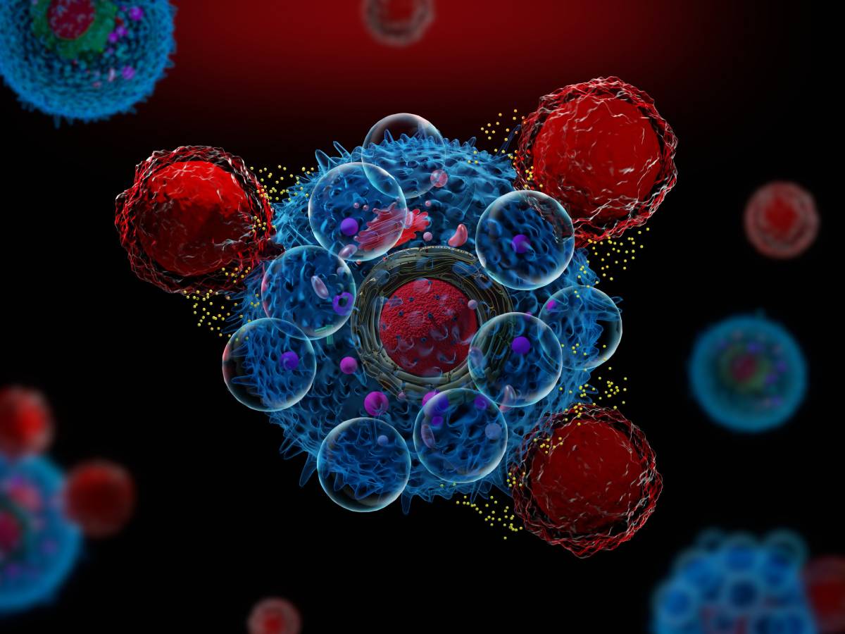
Cytokines are a broad class of cell signaling peptides that exist in the extracellular environment and affect the interactions between cells [1]. Several groups of cytokines, including interleukins and tumor necrosis factors, are known to trigger inflammation in response to infection [2]. Under normal circumstances, a pathogen is bound by pattern recognition receptors, activating pathways that ultimately result in pro-inflammatory cytokine production. These cytokines target the pathogen and recruit leukocytes to the site of infection [3]. However, in the immune reaction known as a “cytokine storm,” certain factors can cause an overproduction of cytokines that may lead to sepsis and other life-threatening conditions. 

The term “cytokine storm” was first used in 1993 to describe an observed immunological response in graft-versus-host disease and has since been used more commonly in reference to infectious diseases [4]. The exact physiology of the cytokine storm is still uncertain, owing largely to the complexity of the cytokine network and the numerous local and systemic effects that these molecules cause. Several classes of growth factors, however, have been strongly implicated in the auto-amplifying phenomenon of the cytokine storm. Specifically, hematopoietic targeted colony stimulating factors that are secreted during the onset of sepsis are known to induce myeloid cell differentiation and proliferation, leading to the increased production of helper T cells and amplified cytokine production [5]. Understanding the genetic basis for the cytokine storm is crucial to deciphering its physiology. Calvano et al. report that white blood cells recently exposed to an endotoxin reveal increased levels of pro-inflammatory gene expression in the 2-4 hour period after exposure and anti-inflammatory cytokine production in the 4-6 hour range; however, the team also found that some 1556 genes are involved in the global response to an endotoxin [6], so this one finding is but one of many genetic insights needed to comprehensively account for the cytokine storm. 

The cytokine storm has received considerable attention in both academic and popular scientific publications in recent months due to its relation to COVID-19. Similar to the pathologies of coronavirus infections SARS and MERS, COVID-19 can lead to increases in pro-inflammatory cytokines and decreases in anti-inflammatory cytokines such as IL-10 [7]. A cytokine storm in response to SARS-CoV-2 infection can cause acute lung damage, which can then lead to acute respiratory distress syndrome, one of the major causes of COVID-19 deaths [8]. Furthermore, as reported by Kaneko et al. in Cell, cytokines may prevent the formation of germinal centers, which could impact the development of long-term immunity [9]. 

Preventing a cytokine storm has thus been a priority for physicians treating COVID-19 patients. Tocilizumab, a drug originally designed for rheumatoid arthritis, has markedly lowered death rates and treatment times for critically ill patients [10]. More controversial has been the use of corticosteroids and the antimalaria drugs chloroquine and hydroxychloroquine in treating COVID; while they may be able to temper the cytokine storm, potential adverse effects may outweigh this benefit [11]. Finally, convalescent plasma therapy has shown positive outcomes: antibodies from a person who has recovered from a coronavirus infection can interact with anti-inflammatory cytokines to block the complementary pro-inflammatory cytokines [12]. More research into the physiology and consequences of the cytokine storm, however, is needed to better treat patients experiencing this dangerous immune reaction. 

References 

1. Zhang, Jun-Ming, and Jianxiong An. “Cytokines, Inflammation and Pain.” International Anesthesiology Clinical, vol. 45, no. 2, 2007, pp. 27–37., www.ncbi.nlm.nih.gov/pmc/articles/PMC2785020/.  

2. Tisoncik, J. R., et al. “Into the Eye of the Cytokine Storm.” Microbiology and Molecular Biology Reviews, vol. 76, no. 1, Mar. 2012, pp. 16–32.  doi:10.1128/MMBR.05015-11

3. Ragab, Dina, et al. “The COVID-19 Cytokine Storm; What We Know So Far.” Frontiers in Immunology, vol. 11, June 2020, p. 1446.  doi:10.3389/fimmu.2020.01446

4. Abhyankar, Sunil, et al. “Interleukin-1 Is A Critical Effector Molecule During Cytokine Dysregulation In Graft Versus Host Disease To Minor Histocompatibility Antigens.” Transplantation, vol. 56, no. 6, Dec. 1993, pp. 1518–22. doi:10.1097/00007890-199312000-00045

5. Chousterman, Benjamin G., et al. “Cytokine Storm and Sepsis Disease Pathogenesis.” Seminars in Immunopathology, vol. 39, no. 5, July 2017, pp. 517–28. doi:10.1007/s00281-017-0639-8

6. Calvano, Steve, and Wenzhong Xiao. “A Network-Based Analysis of Systemic Inflammation in Humans.” Nature, vol. 437, Aug. 2005, pp. 1032–37. 

7. Chen, Jun, and Kanta Subbarao. “The Immunobiology of SARS.” Annual Review of Immunology, vol. 25, no. 1, Apr. 2007, pp. 443–72.  doi:10.1146/annurev.immunol.25.022106.141706

8. Ye, Qing, et al. “The Pathogenesis and Treatment of the `Cytokine Storm’ in COVID-19.” Journal of Infectious Diseases, vol. 80, no. 6, June 2020, pp. 607–13, doi:10.1016/j.jinf.2020.03.037

9. Kaneko, Naoki, et al. “Loss of Bcl-6-Expressing T Follicular Helper Cells and Germinal Centers in COVID-19.” Cell, Aug. 2020, p. S0092867420310679. doi:10.1016/j.cell.2020.08.025

10. “Drug That Calms ‘Cytokine Storm’ Associated with 45% Lower Risk of Dying among COVID-19 Patients on Ventilators.” University of Michiganhttps://labblog.uofmhealth.org/body-work/drug-calms-cytokine-storm-associated-45-lower-risk-of-dying-among-covid-19-patients-on. Accessed 1 Oct. 2020

11. Iannaccone, Giulia, et al. “Weathering the Cytokine Storm in COVID-19: Therapeutic Implications.” Cardiorenal Medicine, vol. 10, no. 5, 2020, pp. 277–87. www.karger.comdoi:10.1159/000509483.  

12. Rojas, Manuel, et al. “Convalescent Plasma in Covid-19: Possible Mechanisms of Action.” Autoimmunity Reviews, vol. 19, no. 7, July 2020, p. 102554. PubMed Centraldoi:10.1016/j.autrev.2020.102554