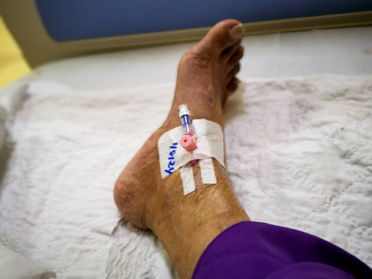
Patient positioning in the perioperative setting is extremely important. While positioning before and during surgery is often discussed, an important aspect of patient care in a procedural setting is patient positioning after surgery and anesthesia. Proper positioning can help prevent various complications such as respiratory distress, circulatory issues, and pressure injuries (Patterson et al., 2024). As anesthesia wears off, patients may experience muscle weakness, shallow breathing, and discomfort. In addition, positioning after surgery and anesthesia must consider the wound site and the risk of nausea and vomiting.

Positioning after surgery and anesthesia can be crucial in patients with postoperative hypoxemia. Postoperative hypoxemia is a condition characterized by low levels of oxygen in the blood. Complications of postoperative hypoxemia include arrhythmias, abnormal blood pressures, and damage to the nervous system (Wang et al., 2024). At the end of a procedure, the supine position is commonly used for extubation. However, in the supine position, the capacity of the lungs is decreased. The semi-recumbent position, where a patient is slightly sitting upwards, is another position that can be used in extubation as it has been shown to increase lung capacity (Wang et al., 2024). In a randomized clinical trial, patients who were positioned in a 30-degree semi-recumbent position during extubation had a lower incidence of hypoxemia (Wang et al., 2024). This may be because diaphragm function improves in the semi-recumbent position compared to the supine position.

Proper circulation after surgery and anesthesia is also an important concern that can be addressed through careful patient positioning. Immobility during and after surgery can increase the risk of blood clots such as deep vein thrombosis. If available, sequential compression devices can reduce risk (SCDs) (Kreutzer et al., 2016). Furthermore, patients should be encouraged to ambulate and move as soon as possible postoperatively. This also helps prevent deep vein thrombosis from occurring (Kreutzer et al., 2016).

The lithotomy position is used during gynecologic, rectal, and urologic surgeries. In this position, patients lie supine with their legs abducted from the midline and knees flexed. However, there are risks with this positioning, like nerve compression. At the end of a procedure, it is important to remove a patient from this position prior to extubation and return them to a supine position to increase lung compliance (Armstrong & Ross, 2022). Following neurosurgery, patients may be placed with their heads elevated to minimize swelling.

Patient positioning after surgery and anesthesia can also affect postoperative nausea and vomiting. Positional changes, especially as a patient is waking up from anesthesia or being transferred, can affect patients who are prone to motion sickness (Patterson et al., 2024). Patients who are sitting rather than supine, even during surgery, have a lower incidence of postoperative nausea and vomiting (Patterson et al., 2024).

Ultimately, positioning after surgery and anesthesia is a critical aspect of recovery. It aids in improving breathing, circulation, and discomfort while reducing the risk of complications including pressure ulcers and respiratory distress. By paying close attention to positioning, healthcare providers can significantly contribute to a smoother and more successful recovery for their patients.

References

Armstrong, Maggie. and Ross A. Moore. “Anatomy, Patient Positioning.” StatPearls, StatPearls Publishing, 31 October 2022.

Wang, Xinghe et al. “Semirecumbent Positioning During Anesthesia Recovery and Postoperative Hypoxemia: A Randomized Clinical Trial.” JAMA network open vol. 7,6 e2416797. 3 Jun. 2024, doi:10.1001/jamanetworkopen.2024.16797

Patterson, Heather et al. “Patient positioning and its impact on perioperative outcomes in children: A narrative review.” Paediatric anaesthesia vol. 34,6 (2024): 507-518. doi:10.1111/pan.14883

Kreutzer, Lindsey et al. “JAMA PATIENT PAGE. Preventing Venous Thromboembolism After Surgery.” JAMA vol. 315,19 (2016): 2136. doi:10.1001/jama.2016.1457

Elective surgery refers to a procedure that is scheduled in advance because it is not performed in an emergency situation. Unlike urgent or emergency surgeries, which are necessary to address acutely life-threatening conditions, elective surgeries are planned at times that are convenient for both the patient and the surgeon. This term encompasses a wide range of procedures, from cosmetic surgeries to important but non-emergency operations such as joint replacements or cataract removal.

Elective surgery is defined by its timing rather than its importance. While the word “elective” might imply that the procedure is optional, many elective surgeries are medically necessary. The main distinction is that these surgeries do not need to be performed immediately and can be scheduled at a later date.

Elective surgeries encompass a wide range of procedures, including cosmetic surgeries like rhinoplasty and breast augmentation, orthopedic surgeries such as hip or knee replacements, and weight-loss surgeries like gastric bypass or sleeve gastrectomy. Eye surgeries, including cataract removal or LASIK, are also common elective procedures, as are certain cardiac surgeries like pacemaker installation or heart valve repair, provided the condition is not immediately life-threatening. General surgeries, such as hernia repairs or gallbladder removal, also typically fall under the category of elective operations 1–3.

Elective surgeries can generally be grouped into two categories based on their necessity and urgency. Medically necessary elective surgeries are procedures that, while not immediately required, are needed to improve a patient’s quality of life or to prevent a condition from worsening. For instance, a person with a painful knee joint due to arthritis might not need immediate surgery, but a knee replacement can significantly improve their ability to move and reduce pain. Similarly, surgeries like removing tumors that are non-cancerous but may cause future complications fall into this category. In contrast, optional or cosmetic elective surgeries are not essential for physical health but are chosen by the patient for personal reasons, often to enhance appearance or correct minor issues. Examples include facelifts, liposuction, or reconstructive procedures following trauma. While these surgeries are elective, they may have a significant psychological impact on a patient, improving self-esteem and confidence 4,5.

Elective surgery offers several benefits, particularly for patients dealing with chronic conditions that affect their quality of life. By addressing issues like joint pain, vision impairment, or weight management, elective surgeries can provide significant physical and emotional relief. However, as with any surgical procedure, there are inherent risks. These risks depend on the type of surgery, the patient’s health, and the complexity of the procedure. Potential risks include infection, bleeding, complications from anesthesia, and postoperative recovery issues. Patients must weigh the potential benefits against the risks and discuss all concerns with their healthcare provider. Because elective surgeries are planned, patients have the advantage of time to consider their options, seek second opinions if necessary, and thoroughly research the procedure and potential outcomes 6–9.

References

1.        Planned surgery (elective surgery) – Better Health Channel. Available at: https://www.betterhealth.vic.gov.au/planned-surgery-elective-surgery.

2.        Types of Surgery — Royal College of Surgeons. Available at: https://www.rcseng.ac.uk/patient-care/having-surgery/types-of-surgery/.

3.        Elective Surgery (for Parents) | Nemours KidsHealth. Available at: https://kidshealth.org/en/parents/elective.html.

4.        Elective Surgeries. Available at: https://www.adena.org/health-focus-blog/detail/adena-health-focus/2022/11/01/elective-surgeries.

5.        Different Types of Surgery | OakBend Medical Center. Available at: https://oakbendmedcenter.org/different-types-of-surgery/.

6.        The Pros and Cons of Elective Surgery: An In-Depth Analysis. Available at: https://www.kirbysurgicalcenter.com/the-pros-and-cons-of-elective-surgery-an-in-depth-analysis.html.

7.        Shaydakov, M. E. & Tuma, F. Operative Risk. (2023).

8.        Operative Risk – StatPearls – NCBI Bookshelf. Available at: https://www.ncbi.nlm.nih.gov/books/NBK532240/. (Accessed: 6th October 2024)

9.        NHS England » New data demonstrates benefits to patients from a range of elective surgeries. Available at: https://www.england.nhs.uk/2016/08/benefit-to-patients/.

Peripheral nerve blocks are widely used in medical practice to provide pain relief during and after surgical procedures, as well as in the management of chronic pain. They involve the injection of anesthetic near a specific nerve or group of nerves to block sensation in a targeted area of the body 1,2. While peripheral nerve blocks are generally considered safe and effective, they are not without potential downsides. Understanding these limitations is crucial for both healthcare providers and patients when considering this pain management option.

One of the downsides of peripheral nerve blocks is the risk of nerve damage. Although uncommon, improper needle placement or accidental puncture of the nerve can lead to temporary or permanent nerve injury. Symptoms of nerve damage may include numbness, tingling, weakness, or even paralysis in the affected area. In some cases, these symptoms may resolve over time, but in others, they can become permanent, leading to chronic pain or loss of function 3.

Infection is a potential complication of any procedure that involves breaking the skin, including peripheral nerve blocks. If proper sterile techniques are not followed, bacteria can be introduced into the injection site, leading to infection. Symptoms of infection may include redness, swelling, warmth, and pain at the injection site. In severe cases, an abscess may form, requiring further medical intervention. Additionally, there is a risk of hematoma formation, especially if the needle punctures a blood vessel. This can lead to localized bleeding and the development of painful swelling or a bruise 3–5.

Patients may experience allergic reactions to the anesthetic agents used in peripheral nerve blocks. While allergic reactions are rare, they can range from mild (such as itching or rash) to severe (such as anaphylaxis, which is a life-threatening condition). It is important for healthcare providers to thoroughly review a patient’s medical history and any known allergies before administering a nerve block to minimize this risk 6.

While peripheral nerve blocks are generally effective, incomplete or failed blocks—when the anesthetic does not fully numb the intended area—come with several downsides including procedural delays, patient discomfort, and re-attempting the block or changing the anesthetic plan. Failed block can result from several factors, including incorrect needle placement, inadequate dosage of anesthetic, or anatomical variations in the patient. A failed block can lead to inadequate pain control during surgery or post-operatively, necessitating the use of additional pain management techniques, such as general anesthesia or systemic analgesics 3.

Systemic toxicity occurs when the anesthetic used in a nerve block is absorbed into the bloodstream at levels that can affect other parts of the body. This can happen if too much anesthetic is used or if the injection inadvertently enters a blood vessel. Symptoms of systemic toxicity can include dizziness, tinnitus (ringing in the ears), seizures, and cardiac arrest. Although rare, this is a serious complication that requires immediate medical attention 3,6,7.

Peripheral nerve blocks are an important tool in pain management, offering significant benefits for patients undergoing surgery or dealing with chronic pain. However, like any medical procedure, peripheral nerve blocks come with risks and potential downsides. Understanding these risks is essential for making informed decisions about their use.

References

1.  Peripheral Nerve Blocks – StatPearls – NCBI Bookshelf. Available at: https://www.ncbi.nlm.nih.gov/books/NBK459210/.

2.  Nerve Block Pros and Cons: – Atlas Pain Specialists. Available at: https://atlaspainspecialists.com/nerve-block-pros-and-cons/.

3. Jeng, C. L., Torrillo, T. M. & Rosenblatt, M. A. Complications of peripheral nerve blocks. Br. J. Anaesth. (2010). doi:10.1093/bja/aeq273

4. Nerve Block: What It Is, Procedure, Side Effects & Types. Available at: https://my.clevelandclinic.org/health/treatments/12090-nerve-blocks.

5. Wiegel, M., Gottschaldt, U., Hennebach, R., Hirschberg, T. & Reske, A. Complications and adverse effects associated with continuous peripheral nerve blocks in orthopedic patients. Anesth. Analg. (2007). doi:10.1213/01.ane.0000261260.69083.f3

6. Local Anesthetic Systemic Toxicity and Allergy to Local Anesthetics | Hadzic’s Peripheral Nerve Blocks and Anatomy for Ultrasound-Guided Regional Anesthesia, 3e | AccessAnesthesiology | McGraw Hill Medical. Available at: https://accessanesthesiology.mhmedical.com/content.aspx?bookid=3074&sectionid=255807496.

7. Wiederhold, B. D., Garmon, E. H., Peterson, E., Stevens, J. B. & O’Rourke, M. C. Nerve Block Anesthesia. StatPearls (2023).

Bariatric surgery is a very effective intervention for individuals with severe obesity, leading to significant and sustained weight loss, improvement in comorbid conditions, and enhanced quality of life 1. Traditionally, bariatric surgery has been performed as an inpatient procedure, requiring patients to stay in the hospital for a few days post-operation. However, advancements in surgical techniques, anesthesia, and postoperative care have turned outpatient bariatric surgery into a viable option for some patients. Patients and their physicians must determine whether inpatient or outpatient bariatric surgery is the best option for them.

Inpatient bariatric surgery involves admitting the patient to the hospital for at least one night post-surgery. Often, this is done for more complex procedures or patients with higher risk profiles. Inpatient surgery allows for enhanced postoperative monitoring and immediate intervention if complications arise, which is particularly important for patients with significant comorbidities or those undergoing more complex procedures. In addition, patients receive comprehensive care, including pain management, nutritional support, and physical therapy, which can improve recovery outcomes. The hospital environment also provides a controlled setting with access to advanced medical equipment and specialists, enhancing overall patient safety 2.

However, inpatient surgery is generally more expensive than outpatient surgery across procedures, including bariatric surgery, due to the extended hospital stay and additional resources required—for high-risk patients though, the increased cost is justified by the enhanced safety and monitoring 3,4. Furthermore, a longer hospital stay can be inconvenient for patients and may increase the risk of hospital-acquired infections 5.

Outpatient bariatric surgery, also known as same-day surgery or ambulatory surgery, involves performing the surgical procedure with the expectation that the patient will be discharged within 24 hours—this is typically reserved for less complex procedures and patients with lower risk profiles.

The most significant advantage of outpatient bariatric surgery is removing the hospital stay 6. Patients can return to the comfort of their homes on the same day 4, which can improve patient psychological well-being and reduce the risk of hospital-acquired infections. In general, patients may find outpatient bariatric surgery thus more convenient than inpatient, as it reduces the disruption to their lives. In addition, outpatient bariatric surgery is generally more cost-effective compared to inpatient surgery due to the reduced need for prolonged hospital stays and associated resources 4.

Not all patients are suitable candidates for outpatient bariatric surgery, as the outpatient setting has reduced access to certain medical services 3. Ideal candidates are therefore typically younger, have a lower body mass index (BMI), and fewer comorbidities—comprehensive preoperative evaluation and risk stratification are essential. Furthermore, ensuring adequate postoperative care is crucial for outpatient procedures, since patients must have access to appropriate follow-up care, including emergency services if complications arise 4.

In conclusion, both outpatient and inpatient bariatric surgery have their unique advantages and considerations. In the end, the choice depends on patient-specific factors, the complexity of the procedure, and the availability of postoperative support.

References

1.          Bariatric surgery – Mayo Clinic. Available at: https://www.mayoclinic.org/tests-procedures/bariatric-surgery/about/pac-20394258.

2.          Is Bariatric Surgery Inpatient or Outpatient?  – Acibadem Health Point International. Available at: https://www.acibademhealthpoint.com/the-is-bariatric-surgery-inpatient-or-outpatient/.

3.          Can the Gastric Sleeve Be Performed as Outpatient Surgery? Available at: https://www.masjax.com/gastric-sleeve-performed-on-an-outpatient-basis/.

4.          How does outpatient gastric sleeve surgery differ from inpatient procedures? Available at: https://gastricsleevelife.com/how-does-outpatient-gastric-sleeve-surgery-differ-from-inpatient-procedures/.

5.        Khan, H. A., Baig, F. K. & Mehboob, R. Nosocomial infections: Epidemiology, prevention, control and surveillance. Asian Pacific Journal of Tropical Biomedicine (2017). doi:10.1016/j.apjtb.2017.01.019

6.        Fortin, S. P., Kalsekar, I., Johnston, S. & Akincigil, A. Comparison of safety and utilization outcomes in inpatient versus outpatient laparoscopic sleeve gastrectomy: a retrospective, cohort study. Surg. Obes. Relat. Dis. (2020). doi:10.1016/j.soard.2020.07.012

Cardiogenic shock is a critical condition that occurs when the heart suddenly cannot pump enough blood to meet the body’s needs. This severe form of cardiac failure is often triggered by a heart attack but can also result from other cardiac issues such as end-stage heart disease, cardiac arrhythmias, or complications from heart surgery. Effective management of cardiogenic shock is crucial, as it is associated with a high mortality rate. Understanding the strategies for timely intervention and continuous monitoring can significantly improve patient outcomes. 

Recognizing the Signs of Cardiogenic Shock 

Early recognition of cardiogenic shock is vital for successful treatment. The primary symptoms include rapid breathing, severe shortness of breath, loss of consciousness, weak pulse, and low blood pressure. Cold and clammy skin, rapid and weak pulse, confusion, and profuse sweating are additional signs that may indicate the onset of this condition. Medical professionals must quickly assess these symptoms to initiate appropriate interventions. 

Immediate Stabilization and Diagnosis 

The initial management of cardiogenic shock involves immediate stabilization of the patient’s condition. This includes ensuring adequate oxygenation and ventilation, providing intravenous fluids to maintain blood pressure, and using medications to support heart function. Once stabilization is achieved, comprehensive diagnostic tests such as echocardiography, electrocardiograms (ECG), and coronary angiography are conducted to determine the underlying cause of the shock. 

Pharmacological Management 

Medications play a critical role in the management of cardiogenic shock. Vasoactive drugs are commonly used to increase cardiac output and improve blood flow. Inotropes like dobutamine and milrinone can enhance heart muscle contractions, whereas vasopressors such as norepinephrine are used to tighten blood vessels and raise blood pressure. Each medication must be carefully selected based on the patient’s specific condition and continuously adjusted according to their response to treatment. 

Mechanical Support Devices 

In cases where pharmacological treatments are not sufficient, mechanical circulatory support devices may be necessary. Devices such as intra-aortic balloon pumps (IABP) and ventricular assist devices (VADs) can provide temporary support to the heart by improving myocardial perfusion and reducing the workload on the heart. These devices are critical in stabilizing patients in severe shock, bridging them either to recovery, a more permanent solution like a heart transplant, or long-term device implantation. 

Addressing the Underlying Cause 

Successful management of cardiogenic shock not only involves symptomatic treatment but also addressing the root cause of the shock. For example, if a heart attack is the trigger, revascularization procedures like percutaneous coronary intervention (PCI) or coronary artery bypass grafting (CABG) may be required to restore blood flow to the heart. Similarly, if a valve disorder or a congenital defect is the cause, surgical or percutaneous repairs may be necessary. 

Continuous Monitoring and Support 

Continuous monitoring is essential in the management of cardiogenic shock. Intensive care units equipped with advanced monitoring technologies provide real-time data on cardiac function, blood pressure, oxygen levels, and other vital parameters. This constant vigilance allows healthcare providers to quickly detect changes in the patient’s condition and adjust treatment protocols accordingly. 

Multidisciplinary Care Team 

Managing cardiogenic shock requires a collaborative approach involving a multidisciplinary team of healthcare professionals. This team typically includes cardiologists, cardiac surgeons, critical care specialists, nurses, and other specialists who work together to provide comprehensive care. Effective communication and coordination among the team members are crucial for ensuring that the patient receives the best possible care. 

Conclusion 

Managing cardiogenic shock effectively requires an integrated approach that combines rapid diagnosis, advanced pharmacological treatments, mechanical support options, and addressing the underlying cause of the condition. With advancements in medical technology and the growing expertise of healthcare professionals, the outcomes for patients with cardiogenic shock have significantly improved. However, continuous education, research, and collaboration across the medical community are essential to further enhance the survival rates and quality of life for these patients. 

Categories
Uncategorized

Time-Outs in the OR

In the high-stakes environment of the operating room (OR), precision, communication, and teamwork are paramount. Among the various protocols implemented to ensure patient safety and surgical success, one common practice is the “time-out”. As defined by the Joint Commission’s Universal Protocol, OR time-outs are deliberate pauses taken before the commencement of surgical procedures to verify critical details and prevent errors. This practice has been widely adopted across healthcare facilities globally, contributing significantly to the reduction of preventable surgical complications.

The practice of time-outs in the OR originated from the need to reduce surgical errors, particularly wrong-site, wrong-procedure, and wrong-patient surgeries. These errors, though rare, can have devastating consequences for patients and healthcare providers alike. The time-out procedure, therefore, serves as a final check to ensure that all team members are aligned and any discrepancies are resolved before the incision is made 1.

A typical time-out involves the entire surgical team and is led by a designated team member, often the surgeon or circulating nurse. The key components verified during a time-out are the patient’s name and date of birth, the specific procedure to be performed, and the correct surgical site. The team ensures as well that the patient’s consent form is accurate and matches the planned procedure and reviews any patient allergies and relevant medical history that could impact the surgery. The team checks that all necessary equipment and supplies are available and functioning. The roles and responsibilities of each team member during the procedure are clarified 2,3.

The implementation of time-outs in the OR has several benefits. First, time-outs foster open communication among the surgical team, ensuring that everyone is on the same page and aware of their roles. This collaborative approach reduces the likelihood of errors and enhances team coordination. Second, by systematically verifying critical information, time-outs significantly reduce the risk of wrong-site, wrong-procedure, and wrong-patient surgeries. They also help identify and rectify any inconsistencies or missing information before the procedure begins. Finally, adhering to time-out protocols helps healthcare facilities comply with regulatory standards and accreditation requirements, such as those set by the Joint Commission. This compliance is important for maintaining a facility’s reputation and operational status 1,3.

Despite their benefits, OR time-outs can have a number of challenges. Some team members may view them as redundant or time-consuming, particularly in high-volume surgical settings. Additionally, the effectiveness of a time-out depends on the commitment and cooperation of the entire surgical team. To address these challenges, ongoing education and training, as well as refining protocols, can improve the use of time-outs 3,4.

Time-outs in the OR are a simple yet powerful tool in the quest for surgical excellence. By fostering meticulous attention to detail and encouraging robust team communication, time-outs play a critical role in preventing errors and enhancing patient safety.

References

1.        Enhancing Patient Safety: Understanding Timeouts in the Operating Room | Skytron, LLC. Available at: https://www.skytron.com/articles/enhancing-patient-safety-understanding-timeouts-in-the-operating-room/. (Accessed: 20th June 2024)

2.        Li, G. et al. Postoperative Pulmonary Complications’ Association with Sugammadex versus Neostigmine: A Retrospective Registry Analysis. Anesthesiology (2021). doi:10.1097/ALN.0000000000003735

3.        Papadakis, M., Meiwandi, A. & Grzybowski, A. The WHO safer surgery checklist time out procedure revisited: Strategies to optimise compliance and safety. International Journal of Surgery (2019). doi:10.1016/j.ijsu.2019.07.006

4.        Article | Outpatient Surgery Magazine. Available at: https://www.aorn.org/outpatient-surgery/article/2018-December-your-attention-please-time-for-a-time-out. (Accessed: 20th June 2024)

Post dural puncture headache (PDPH), also referred to as post lumbar puncture headache, is a frequent complication arising from diagnostic lumbar puncture. It can also occur after spinal anesthesia or, more commonly, from accidental dural puncture during an epidural catheter placement attempt. The headache is typically positional, becoming worse when standing and improving when lying down. PDPH is often associated with neck stiffness, sensitivity to light (photophobia), nausea, or subjective hearing symptoms. In some cases, symptoms can be debilitating, requiring medical intervention. One such intervention for PDPH is an invasive treatment known as an epidural blood patch (EBP).

The exact cause of headache following a dural puncture is not entirely understood, but it is believed to be related to the leakage of cerebrospinal fluid (CSF) through the dural hole made by the needle. If CSF leaks faster than it is produced, low CSF pressure can occur, which is more pronounced at the level of the brain when in an upright position. However, not all patients with PDPH have low CSF pressure, and not everyone with a significant CSF leak develops a headache. The incidence of PDPH varies greatly, depending on patient-specific and procedural risk factors. After spinal anesthesia, it is between 3% and 9%, depending on the type and size of the needle used. After lumbar puncture, it occurs in about 11% of cases.

The treatment of PDPH depends on the severity of the headache and its effect on the patient’s ability to function. Patients with PDPH who cannot tolerate sitting or standing, who are unable to carry out daily activities, or whose headache does not respond to a short trial of conservative measures are considered to have moderate to severe PDPH. These patients should be offered an epidural blood patch, which may provide lasting symptomatic relief.

An EBP is a procedure where a small amount of the patient’s own blood is injected into the epidural space to seal a CSF leak. Epidural blood patch is regarded as the definitive treatment for PDPH. A systematic review by Boonmak et al., published in 2010, found that EBP reduced the duration and intensity of post-dural puncture headache compared to both conservative treatment and sham procedures. EBP typically provides immediate relief. The success rate after the first EBP is between 65% and 98%, with a similar success rate for a second EBP if needed.

Similar to epidural anesthesia, an epidural blood patch is contraindicated for patients with coagulopathy, those on anticoagulants, and patients with systemic infections or infections at the epidural needle insertion site. The EBP procedure involves injecting the patient’s blood into the epidural space using an epidural needle. The needle is inserted as it would be for epidural anesthesia, using the loss of resistance to saline technique to locate the epidural space without inserting a catheter. Once the epidural space is identified, a second operator draws a syringe of the patient’s venous blood using aseptic technique, which is then slowly injected through the epidural needle. Headache symptoms typically improve within seconds to minutes after the procedure, although a temporary sensation of “fullness” in the back is common. After the EBP is completed, the patient is advised to lie flat for one to two hours with minimal movement. Following this period, the patient can stand and resume normal activities.

References

DelPizzo K, Cheng J, Dong N, Edmonds CR, Kahn RL, Fields KG, Curren J, Rotundo V, Zayas VM. Post-Dural Puncture Headache is Uncommon in Young Ambulatory Surgery Patients. HSS J. 2017 Jul;13(2):146-151. doi: 10.1007/s11420-017-9541-0. Epub 2017 Mar 16. PMID: 28690464; PMCID: PMC5481263.

Choi PT, Galinski SE, Takeuchi L, Lucas S, Tamayo C, Jadad AR. PDPH is a common complication of neuraxial blockade in parturients: a meta-analysis of obstetrical studies. Can J Anaesth. 2003 May;50(5):460-9. doi: 10.1007/BF03021057. PMID: 12734154.

Vallejo MC, Mandell GL, Sabo DP, Ramanathan S. Postdural puncture headache: a randomized comparison of five spinal needles in obstetric patients. Anesth Analg. 2000 Oct;91(4):916-20. doi: 10.1097/00000539-200010000-00027. PMID: 11004048.

Boonmak P, Boonmak S. Epidural blood patching for preventing and treating post-dural puncture headache. Cochrane Database Syst Rev. 2010 Jan 20;(1):CD001791. doi: 10.1002/14651858.CD001791.pub2. Update in: Cochrane Database Syst Rev. 2013;11:CD001791. PMID: 20091522.

Abouleish E, Vega S, Blendinger I, Tio TO. Long-term follow-up of epidural blood patch. Anesth Analg. 1975 Jul-Aug;54(4):459-63. doi: 10.1213/00000539-197554040-00012. PMID: 125053.

Paech MJ, Doherty DA, Christmas T, Wong CA; Epidural Blood Patch Trial Group. The volume of blood for epidural blood patch in obstetrics: a randomized, blinded clinical trial. Anesth Analg. 2011 Jul;113(1):126-33. doi: 10.1213/ANE.0b013e318218204d. Epub 2011 May 19. PMID: 21596867.

Banks S, Paech M, Gurrin L. An audit of epidural blood patch after accidental dural puncture with a Tuohy needle in obstetric patients. Int J Obstet Anesth. 2001 Jul;10(3):172-6. doi: 10.1054/ijoa.2000.0826. PMID: 15321606.

Total shoulder arthroplasty (TSA) is a surgical procedure that aims to provide relief and improved function for patients with severe shoulder pathologies, such as osteoarthritis, rheumatoid arthritis, or post-traumatic arthritis (1). To ensure patient comfort and improve postoperative recovery, effective regional anesthesia techniques are essential during total shoulder replacement surgery. These techniques not only provide pain relief, but also reduce the need for systemic opioids and their associated side effects.

The shoulder is innervated primarily by the brachial plexus, a complex network of nerves that arise from the cervical and upper thoracic spinal nerves (C5-T1). The brachial plexus branches into several key nerves that are targets in TSA, including the suprascapular, axillary, and lateral pectoral nerves (1). Effective anesthesia for total shoulder replacement surgery involves targeting these nerves to ensure both pain control and muscle relaxation.

There are several nerve block techniques used in TSA, each targeting different nerves of the brachial plexus to optimize pain control. One of the primary blocks used is the interscalene brachial plexus block, which effectively numbs the nerves responsible for transmitting most shoulder pain. The interscalene block provides excellent analgesia but carries risks such as diaphragm paralysis due to phrenic nerve involvement, making appropriate patient selection critical (2). Another technique, the suprascapular nerve block, specifically targets the suprascapular nerve, which innervates approximately 70% of the shoulder joint. This block can be used alone or in conjunction with the interscalene block to improve pain control, which is especially beneficial in patients who are at significant risk for diaphragmatic paralysis. The axillary nerve block is another technique that is often used in combination with the aforementioned blocks to provide comprehensive coverage of the sensory nerves of the shoulder (2). This block is typically performed under ultrasound guidance to increase accuracy and reduce complications.

Advances in ultrasound technology have significantly improved the safety and efficacy of these nerve blocks by allowing anesthesiologists to visualize the nerve and surrounding tissue during the injection of anesthetic agents. The use of ultrasound has been shown to reduce the incidence of complications and improve the quality of the nerve block.

When selecting anesthetic agents, a variety of medications can be used for regional anesthesia for total shoulder replacement surgery. Bupivacaine and ropivacaine are commonly used local anesthetics due to their long duration of action, which is beneficial for postoperative pain management. These agents can be administered alone or in combination with adjuvants such as epinephrine, which prolongs the duration of the block and reduces systemic absorption, thereby minimizing toxicity. Recently, the use of liposomal bupivacaine, an extended-release formulation, has gained popularity (1). This agent provides prolonged postoperative analgesia, which can significantly improve patient comfort and reduce the need for additional opioid analgesia. In addition, adjuvants such as dexmedetomidine or dexamethasone are increasingly being used to prolong the effects of nerve blocks (3). These adjuvants, when added to traditional local anesthetics, have been shown to prolong the duration of analgesia without significant additional side effects.

The anesthesia approach for total shoulder replacement surgery should be individualized based on the patient’s specific medical condition, surgical procedure, and potential risk factors for complications. The integration of ultrasound-guided nerve blocks and the strategic use of long-acting local anesthetics and adjuvants can significantly improve the quality of postoperative pain management, thereby enhancing recovery and patient satisfaction in total shoulder arthroplasty.

References

  1. Fredrickson MJ, Krishnan S, Chen CY. Postoperative analgesia for shoulder surgery: a critical appraisal and review of current techniques. Anaesthesia. 2010;65(6):608-624. doi:10.1111/j.1365-2044.2009.06231.x
  2. Bishop JY, Sprague M, Gelber J, et al. Interscalene regional anesthesia for shoulder surgery. J Bone Joint Surg Am. 2005;87(5):974-979. doi:10.2106/JBJS.D.02003
  3. Vorobeichik L, Brull R, Abdallah FW. Evidence basis for using perineural dexmedetomidine to enhance the quality of brachial plexus nerve blocks: a systematic review and meta-analysis of randomized controlled trials. Br J Anaesth. 2017;118(2):167-181. doi:10.1093/bja/aew411
Categories
Uncategorized

Point-of-Care Testing

In fast-paced medical environments, quick and accurate diagnostic information can be critical for guiding treatment. Point-of-care testing (POCT) is a growing concept in healthcare, especially in the context of anesthesia and surgery, that emphasizes real-time diagnostic results at the patient’s bedside.

POCT involves performing diagnostic tests at the “point of care” rather than sending samples to a centralized laboratory. This approach offers several advantages, including rapid turnaround time, enhanced workflow efficiency, and improved patient outcomes. By providing timely insights into a patient’s condition, POCT enables clinicians to make informed decisions promptly and gather more information for time-sensitive decisions 1.

In the context of the operating room (OR), where speed and efficiency are particularly important, point-of-care testing has revolutionized the way surgical teams manage patient care. One of the most common applications of POCT in the OR is the measurement of blood gases and electrolytes. Monitoring arterial blood gas levels during surgery is crucial for assessing respiratory function, acid-base balance, and oxygenation status in critically ill patients. With POCT devices, doctors can obtain arterial blood gas results within minutes, allowing for prompt adjustments in ventilation and oxygen therapy to optimize patient outcomes 2.

Moreover, POCT plays a vital role in monitoring coagulation parameters during surgical procedures, especially in patients receiving anticoagulant therapy or undergoing major surgeries. Devices such as point-of-care coagulation analyzers enable rapid testing of clotting function, allowing surgeons to make timely decisions regarding blood transfusions, hemostatic interventions, and the management of bleeding complications 2.

Another area where point-of-care testing is making a significant impact in the OR is in the detection of infectious diseases. Rapid diagnostic tests for pathogens such as methicillin-resistant Staphylococcus aureus (MRSA) and Clostridioides difficile (C. diff) enable early identification of healthcare-associated infections to guide appropriate antimicrobial therapy and infection control measures. By identifying infectious agents quickly, POCT helps reduce the risk of postoperative complications and hospital-acquired infections, ultimately improving patient safety and outcomes 1.

Furthermore, POCT facilitates the monitoring of glucose levels in diabetic patients undergoing surgery, ensuring tight glycemic control and minimizing the risk of perioperative complications such as hyperglycemia and hypoglycemia. Portable glucose meters allow surgical teams to perform frequent blood glucose measurements intraoperatively, guiding insulin administration and fluid management strategies to maintain optimal metabolic balance 1,3.

POCT offers clinical and clear logistical advantages. By eliminating the need to transport samples to a central laboratory and wait for results, it reduces turnaround times and streamlines the decision-making process during surgery. This not only enhances workflow efficiency but also conserves valuable resources and reduces healthcare costs associated with traditional laboratory testing 4.

However, point-of-care testing in the OR also has some disadvantages. Quality control, operator training, and compliance with regulatory standards are essential to ensuring the accuracy and reliability of results. Moreover, interoperability and integration with electronic medical records systems are crucial for data management and documentation in the perioperative setting 4.

Overall, point-of-care testing is advancing healthcare delivery by providing rapid and reliable diagnostic information that empowers surgical teams to make informed decisions and optimize patient care. As technology continues to evolve, the integration of POCT into routine practice is poised to further enhance patient safety, outcomes, and overall experience.

References

  1. Point-of-Care Testing – StatPearls – NCBI Bookshelf. Available at: https://www.ncbi.nlm.nih.gov/books/NBK592387/. (Accessed: 17th March 2024)
  2. Point-of-Care Testing – Testing.com. Available at: https://www.testing.com/articles/point-of-care-testing/. (Accessed: 17th March 2024)
  3. Point of Care Testing | Sight Diagnostics. Available at: https://sightdx.com/en/knowledge-center/point-of-care-testing. (Accessed: 17th March 2024)
  4. Point of Care Testing (POCT). Available at: https://www.leedsth.nhs.uk/a-z-of-services/pathology/poct/. (Accessed: 17th March 2024)

High or total spinal anesthesia refers to the administration of local anesthetic agents at a level near the upper thoracic or cervical spine, resulting in a block of sensation and motor function in the body (1). This type of anesthesia is typically used for surgeries involving the upper abdomen, chest, or neck, providing a more extensive area of numbness compared to lower spinal anesthesia. Unfortunately, high spinal anesthesia can cause respiratory complications due to its effects on the nerves that control the diaphragm and other respiratory muscles. Respiratory complications associated with high spinal anesthesia include difficulty breathing, decreased lung function, and in severe cases, respiratory failure (2). Understanding these effects is critical for anesthesiologists and other anesthesia providers to ensure patient safety and optimize outcomes during surgical procedures.

It is important for anesthesia providers to carefully monitor the patient’s oxygen saturation levels, end-tidal carbon dioxide levels, and respiratory effort to identify potential complications early. In addition, maintaining proper patient positioning during surgery can help prevent respiratory problems associated with high spinal anesthesia (1). Overall, a proactive approach to managing respiratory changes during surgery plays a critical role in ensuring patient comfort and successful postoperative outcomes. This proactive approach includes closely monitoring the patient’s vital signs, such as heart rate and blood pressure, and assessing for signs of respiratory distress, such as increased work of breathing or cyanosis. Surgeons may need to modify their approach or technique to minimize the risk of respiratory complications, while nurses play a key role in monitoring the patient’s response to interventions and providing support as needed (2).

For example, if a patient undergoing surgery begins to show signs of respiratory distress, such as shallow breathing or decreased oxygen saturation, the anesthesia provider may need to increase ventilatory support or administer supplemental oxygen. The surgeon may need to adjust his or her technique to reduce pressure on the patient’s chest or airway, while the nursing staff provides close assistance as needed to ensure optimal respiratory function (3). Effective communication and teamwork are essential to ensure that all members of the healthcare team work together to prioritize the patient’s respiratory health and overall well-being.

In the postoperative period after high spinal anesthesia, it is also critical that providers closely monitor the patient’s vital signs and oxygen saturation to detect early signs of respiratory distress, as risk does not disappear immediately. Prompt intervention and collaboration between the surgical team, nursing staff, and respiratory therapists can help prevent complications and improve the patient’s recovery process (3).  In addition, patient education on deep breathing exercises and early mobilization can play an important role in promoting optimal respiratory function and preventing postoperative complications. This comprehensive approach to respiratory care may also include appropriate pain management to promote deep breathing and coughing, as well as implementing early mobilization strategies to prevent atelectasis and pneumonia (1). By implementing a multidisciplinary approach and emphasizing the importance of respiratory care, healthcare providers can improve patient outcomes and ensure a smooth recovery process after surgery.

References

  1. Aronson JK. Anesthetics, local. In: Meyler’s Side Effects of Drugs. 16th ed. Elsevier; 2015
  2. Sharma J, Bala R, Kumar R, Malhan S. Respiratory arrest after spinal anaesthesia: a conundrum for anesthesiologists. Research and Opinion in Anesthesia and Intensive Care. 2019;6(3):377. doi:10.4103/roaic.roaic_51_18
  3. Foster LA, Deutz CK, Hutchins JL, Allen JA. Total spinal and brainstem anesthesia as complication of paravertebral ropivacaine administration. Neurol Clin Pract. 2017;7(5):430-432. doi:10.1212/CPJ.0000000000000355