
For Informational Purposes Only
Diaphragm Weakness in Patients Undergoing Anesthesia and Surgery
Disclaimer: This article is intended solely for informational and educational purposes only. It does not constitute medical advice.
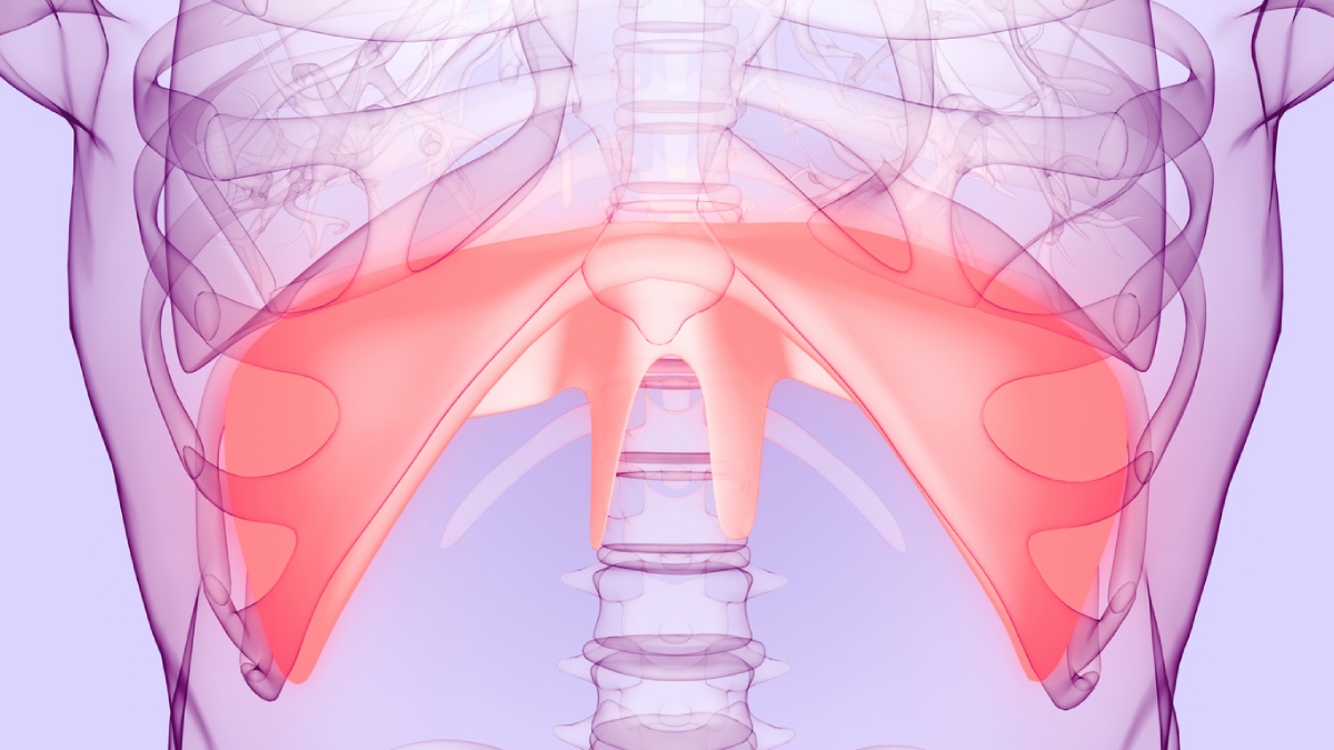
The diaphragm is the primary muscle of inspiration, contributing approximately 60%–80% of normal breathing. Diaphragm weakness can therefore pose a significant concern during anesthesia and surgery, particularly when respiratory function is already compromised. This weakness may result from impaired muscle contractility or injury to the phrenic nerve, which controls diaphragmatic movement. Although unilateral dysfunction is often compensated for by the unaffected hemidiaphragm and other respiratory muscles, bilateral involvement can cause substantial breathing difficulties. Even unilateral weakness may become clinically significant in patients with chronic obstructive pulmonary disease (COPD), obesity, or obstructive sleep apnea, as these conditions can limit respiratory reserve and make it more difficult to compensate for reduced diaphragmatic function (1).
General anesthesia can impair diaphragmatic function through the effects of anesthetic medications and mechanical ventilation. Sedatives and opioids suppress respiratory drive, while neuromuscular blocking agents inhibit diaphragmatic contraction. During controlled mechanical ventilation, reduced muscle activity can lead to ventilator-induced diaphragm dysfunction, which is characterized by muscle fiber atrophy and impaired contractility. These changes may begin within 12–24 hours and become more pronounced with prolonged ventilatory support (1). However, excessive diaphragmatic inactivity is not the only concern, as insufficient ventilatory assistance can increase inspiratory effort and place additional strain on the muscle. Maintaining an appropriate level of diaphragmatic activity is therefore important to minimize injury from both excessive unloading and increased respiratory effort (2).
Regional anesthesia and cardiothoracic surgery may also lead to diaphragm weakness through impairment of the phrenic nerve, which controls diaphragmatic contraction. Interscalene brachial plexus blockade, commonly used for shoulder surgery, can temporarily block the phrenic nerve when local anesthetic spreads beyond the intended injection site. This may cause paralysis of the affected hemidiaphragm, reducing respiratory capacity and potentially compromising ventilation in patients with limited pulmonary reserve (3). During cardiac surgery, the phrenic nerve is vulnerable to injury because of its close proximity to the heart. Surgical manipulation or exposure to cold cardioplegia can damage the nerve, resulting in diaphragmatic weakness that may persist after surgery and interfere with respiratory recovery (4).
Early recognition of diaphragmatic dysfunction is particularly important in patients who experience persistent respiratory difficulties following surgery. Diaphragm ultrasonography provides a noninvasive bedside method for assessing diaphragmatic function through measurements of excursion, muscle thickness, and thickening during inspiration. These measurements can help identify impaired diaphragmatic activity and monitor changes throughout the perioperative period. However, in mechanically ventilated patients, positive-pressure ventilation produces passive diaphragm movement without active muscle contraction, limiting the reliability of diaphragmatic excursion as an indicator of muscle function (1).
It is important to identify patients at increased risk of respiratory impairment through preoperative assessment of respiratory function and underlying conditions. This evaluation can help guide anesthetic selection, particularly when regional techniques may affect the phrenic nerve. During mechanical ventilation, maintaining appropriate diaphragmatic activity by avoiding both excessive ventilatory assistance and increased inspiratory effort may help limit muscle injury. Postoperative management should emphasize adequate reversal of neuromuscular blockade, early extubation when safe, and respiratory support when diaphragmatic weakness persists (1).
References
- Hu J, Guo R, Li H, Wen H, Wang Y. Perioperative Diaphragm Dysfunction. J Clin Med. 2024;13(2):519. Published 2024 Jan 17. doi:10.3390/jcm13020519
- Goligher EC, Dres M, Fan E, et al. Mechanical Ventilation-induced Diaphragm Atrophy Strongly Impacts Clinical Outcomes. Am J Respir Crit Care Med. 2018;197(2):204-213. doi:10.1164/rccm.201703-0536OC
- Urmey WF, McDonald M. Hemidiaphragmatic paresis during interscalene brachial plexus block: effects on pulmonary function and chest wall mechanics. Anesth Analg. 1992;74(3):352-357. doi:10.1213/00000539-199203000-00006
- Laghlam D, Naudin C, Srour A, et al. Persistent diaphragm dysfunction after cardiac surgery is associated with adverse respiratory outcomes: a prospective observational ultrasound study. Can J Anaesth. 2023;70(2):228-236. doi:10.1007/s12630-022-02360-8